Zhi-Peng Guo , Lei Chen , Li-Rong Tang , Yue Gao , Miao Qu , Lihong Wang , Chun-Hong Liu
{"title":"非典型重度抑郁症与典型重度抑郁症之间的眶额活动和连接性差异","authors":"Zhi-Peng Guo , Lei Chen , Li-Rong Tang , Yue Gao , Miao Qu , Lihong Wang , Chun-Hong Liu","doi":"10.1016/j.nicl.2024.103717","DOIUrl":null,"url":null,"abstract":"<div><h3>Objective</h3><div>Atypical major depressive disorder (MDD) is a distinct subtype of MDD, characterized by increased appetite and/or weight gain, excessive sleep, leaden paralysis, and interpersonal rejection sensitivity. Delineating different neural circuits associated with atypical and typical MDD would better inform clinical personalized interventions.</div></div><div><h3>Methods</h3><div>Using resting-state fMRI, we investigated the voxel-level regional homogeneity (ReHo) and functional connectivity (FC) in 55 patients with atypical MDD, 51 patients with typical MDD, and 49 healthy controls (HCs). Support vector machine (SVM) approaches were applied to examine the validity of the findings in distinguishing the two types of MDD.</div></div><div><h3>Results</h3><div>Compared to patients with typical MDD and HCs, patients with atypical MDD had increased ReHo values in the right lateral orbitofrontal cortex (OFC) and enhanced FC between the right lateral OFC and right dorsolateral prefrontal cortex (dlPFC), and between the right striatum and left OFC. The ReHo in the right lateral OFC and the significant FCs found were significantly correlated with body mass index (BMI) in all groups of participants with MDD. The connectivity of the right striatum and left OFC was positively correlated with the retardation scores in the atypical MDD group. Using the ReHo of the right lateral OFC as a feature, we achieved 76.42% accuracy to differentiate atypical MDD from typical MDD.</div></div><div><h3>Conclusion</h3><div>Our findings show that atypical MDD might be associated with altered OFC activity and connectivity. Furthermore, our findings highlight the key role of lateral OFC in atypical MDD, which may provide valuable information for future personalized interventions.</div></div>","PeriodicalId":54359,"journal":{"name":"Neuroimage-Clinical","volume":"45 ","pages":"Article 103717"},"PeriodicalIF":3.4000,"publicationDate":"2024-11-26","publicationTypes":"Journal Article","fieldsOfStudy":null,"isOpenAccess":false,"openAccessPdf":"","citationCount":"0","resultStr":"{\"title\":\"The differential orbitofrontal activity and connectivity between atypical and typical major depressive disorder\",\"authors\":\"Zhi-Peng Guo , Lei Chen , Li-Rong Tang , Yue Gao , Miao Qu , Lihong Wang , Chun-Hong Liu\",\"doi\":\"10.1016/j.nicl.2024.103717\",\"DOIUrl\":null,\"url\":null,\"abstract\":\"<div><h3>Objective</h3><div>Atypical major depressive disorder (MDD) is a distinct subtype of MDD, characterized by increased appetite and/or weight gain, excessive sleep, leaden paralysis, and interpersonal rejection sensitivity. Delineating different neural circuits associated with atypical and typical MDD would better inform clinical personalized interventions.</div></div><div><h3>Methods</h3><div>Using resting-state fMRI, we investigated the voxel-level regional homogeneity (ReHo) and functional connectivity (FC) in 55 patients with atypical MDD, 51 patients with typical MDD, and 49 healthy controls (HCs). Support vector machine (SVM) approaches were applied to examine the validity of the findings in distinguishing the two types of MDD.</div></div><div><h3>Results</h3><div>Compared to patients with typical MDD and HCs, patients with atypical MDD had increased ReHo values in the right lateral orbitofrontal cortex (OFC) and enhanced FC between the right lateral OFC and right dorsolateral prefrontal cortex (dlPFC), and between the right striatum and left OFC. The ReHo in the right lateral OFC and the significant FCs found were significantly correlated with body mass index (BMI) in all groups of participants with MDD. The connectivity of the right striatum and left OFC was positively correlated with the retardation scores in the atypical MDD group. Using the ReHo of the right lateral OFC as a feature, we achieved 76.42% accuracy to differentiate atypical MDD from typical MDD.</div></div><div><h3>Conclusion</h3><div>Our findings show that atypical MDD might be associated with altered OFC activity and connectivity. Furthermore, our findings highlight the key role of lateral OFC in atypical MDD, which may provide valuable information for future personalized interventions.</div></div>\",\"PeriodicalId\":54359,\"journal\":{\"name\":\"Neuroimage-Clinical\",\"volume\":\"45 \",\"pages\":\"Article 103717\"},\"PeriodicalIF\":3.4000,\"publicationDate\":\"2024-11-26\",\"publicationTypes\":\"Journal Article\",\"fieldsOfStudy\":null,\"isOpenAccess\":false,\"openAccessPdf\":\"\",\"citationCount\":\"0\",\"resultStr\":null,\"platform\":\"Semanticscholar\",\"paperid\":null,\"PeriodicalName\":\"Neuroimage-Clinical\",\"FirstCategoryId\":\"3\",\"ListUrlMain\":\"https://www.sciencedirect.com/science/article/pii/S221315822400158X\",\"RegionNum\":2,\"RegionCategory\":\"医学\",\"ArticlePicture\":[],\"TitleCN\":null,\"AbstractTextCN\":null,\"PMCID\":null,\"EPubDate\":\"\",\"PubModel\":\"\",\"JCR\":\"Q2\",\"JCRName\":\"NEUROIMAGING\",\"Score\":null,\"Total\":0}","platform":"Semanticscholar","paperid":null,"PeriodicalName":"Neuroimage-Clinical","FirstCategoryId":"3","ListUrlMain":"https://www.sciencedirect.com/science/article/pii/S221315822400158X","RegionNum":2,"RegionCategory":"医学","ArticlePicture":[],"TitleCN":null,"AbstractTextCN":null,"PMCID":null,"EPubDate":"","PubModel":"","JCR":"Q2","JCRName":"NEUROIMAGING","Score":null,"Total":0}
The differential orbitofrontal activity and connectivity between atypical and typical major depressive disorder
Objective
Atypical major depressive disorder (MDD) is a distinct subtype of MDD, characterized by increased appetite and/or weight gain, excessive sleep, leaden paralysis, and interpersonal rejection sensitivity. Delineating different neural circuits associated with atypical and typical MDD would better inform clinical personalized interventions.
Methods
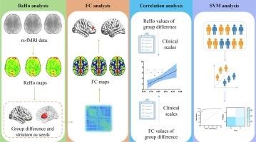
Using resting-state fMRI, we investigated the voxel-level regional homogeneity (ReHo) and functional connectivity (FC) in 55 patients with atypical MDD, 51 patients with typical MDD, and 49 healthy controls (HCs). Support vector machine (SVM) approaches were applied to examine the validity of the findings in distinguishing the two types of MDD.
Results
Compared to patients with typical MDD and HCs, patients with atypical MDD had increased ReHo values in the right lateral orbitofrontal cortex (OFC) and enhanced FC between the right lateral OFC and right dorsolateral prefrontal cortex (dlPFC), and between the right striatum and left OFC. The ReHo in the right lateral OFC and the significant FCs found were significantly correlated with body mass index (BMI) in all groups of participants with MDD. The connectivity of the right striatum and left OFC was positively correlated with the retardation scores in the atypical MDD group. Using the ReHo of the right lateral OFC as a feature, we achieved 76.42% accuracy to differentiate atypical MDD from typical MDD.
Conclusion
Our findings show that atypical MDD might be associated with altered OFC activity and connectivity. Furthermore, our findings highlight the key role of lateral OFC in atypical MDD, which may provide valuable information for future personalized interventions.
期刊介绍:
NeuroImage: Clinical, a journal of diseases, disorders and syndromes involving the Nervous System, provides a vehicle for communicating important advances in the study of abnormal structure-function relationships of the human nervous system based on imaging.
The focus of NeuroImage: Clinical is on defining changes to the brain associated with primary neurologic and psychiatric diseases and disorders of the nervous system as well as behavioral syndromes and developmental conditions. The main criterion for judging papers is the extent of scientific advancement in the understanding of the pathophysiologic mechanisms of diseases and disorders, in identification of functional models that link clinical signs and symptoms with brain function and in the creation of image based tools applicable to a broad range of clinical needs including diagnosis, monitoring and tracking of illness, predicting therapeutic response and development of new treatments. Papers dealing with structure and function in animal models will also be considered if they reveal mechanisms that can be readily translated to human conditions.