四种颞下颌关节骨关节炎小鼠模型下颌骨髁状突的组织病理学特征。

IF 2.2 4区 医学 Q2 DENTISTRY, ORAL SURGERY & MEDICINE
Ludan Xing , Xin Qi , Jiayan Wu , Yuan Liu , Liyuan Zhang , Beizhan Jiang
{"title":"四种颞下颌关节骨关节炎小鼠模型下颌骨髁状突的组织病理学特征。","authors":"Ludan Xing ,&nbsp;Xin Qi ,&nbsp;Jiayan Wu ,&nbsp;Yuan Liu ,&nbsp;Liyuan Zhang ,&nbsp;Beizhan Jiang","doi":"10.1016/j.archoralbio.2024.106129","DOIUrl":null,"url":null,"abstract":"<div><h3>Objective</h3><div>Temporomandibular joint osteoarthritis (TMJOA) has been modeled in different ways with a lack of uniformity. We aimed to investigate four TMJOA mouse models and assess histopathological changes in condyles, which could assist in the selection of animal models in further TMJOA-related studies.</div></div><div><h3>Design</h3><div>Four TMJOA mouse models were established, including unilateral hyperocclusion, discectomy, monosodium iodoacetate injection and aged model. Bilateral condyles were collected at different time points. The condylar alterations were analyzed by Micro-CT, Hematoxylin and eosin staining, Toluidine blue staining, Safranin O staining, Trap staining, immunofluorescence staining, immunohistochemistry staining and quantitative polymerase chain reaction.</div></div><div><h3>Results</h3><div>Radiographic and histopathological analysis indicated that all four methods could cause condylar degeneration successfully. Differences in morphologic and histologic changes were found among four models. The hyperocclusion model was time-dependent and the lesions got worse over time. Discectomy model presented obvious damage of cartilage and subchondral bone. Injected model showed severe inflammation and chondrocyte hypertrophy. The aged model was characterized by decreased of proteoglycan and osteolysis.</div></div><div><h3>Conclusions</h3><div>The four methods had different characteristics and applicability. The harvest time affected the degree of cartilage degradation. Hyperocclusion was suited to explore the early-stage of TMJOA. Discectomy present advantages in investigating the long-term restoration of cartilage and subchondral bone. Monosodium iodoacetate-injection was appropriate for screening the agents for inflammatory relief. The aged model more naturally facilitated discovering underlying mechanisms in primary TMJOA. Unilateral modeling methods could initiate contralateral condylar alterations. The TMJOA models should be selected based on experimental requirements and applicability of each model.</div></div>","PeriodicalId":8288,"journal":{"name":"Archives of oral biology","volume":"170 ","pages":"Article 106129"},"PeriodicalIF":2.2000,"publicationDate":"2024-11-14","publicationTypes":"Journal Article","fieldsOfStudy":null,"isOpenAccess":false,"openAccessPdf":"","citationCount":"0","resultStr":"{\"title\":\"Histopathological characterization of mandibular condyles in four temporomandibular joint osteoarthritis mouse models\",\"authors\":\"Ludan Xing ,&nbsp;Xin Qi ,&nbsp;Jiayan Wu ,&nbsp;Yuan Liu ,&nbsp;Liyuan Zhang ,&nbsp;Beizhan Jiang\",\"doi\":\"10.1016/j.archoralbio.2024.106129\",\"DOIUrl\":null,\"url\":null,\"abstract\":\"<div><h3>Objective</h3><div>Temporomandibular joint osteoarthritis (TMJOA) has been modeled in different ways with a lack of uniformity. We aimed to investigate four TMJOA mouse models and assess histopathological changes in condyles, which could assist in the selection of animal models in further TMJOA-related studies.</div></div><div><h3>Design</h3><div>Four TMJOA mouse models were established, including unilateral hyperocclusion, discectomy, monosodium iodoacetate injection and aged model. Bilateral condyles were collected at different time points. The condylar alterations were analyzed by Micro-CT, Hematoxylin and eosin staining, Toluidine blue staining, Safranin O staining, Trap staining, immunofluorescence staining, immunohistochemistry staining and quantitative polymerase chain reaction.</div></div><div><h3>Results</h3><div>Radiographic and histopathological analysis indicated that all four methods could cause condylar degeneration successfully. Differences in morphologic and histologic changes were found among four models. The hyperocclusion model was time-dependent and the lesions got worse over time. Discectomy model presented obvious damage of cartilage and subchondral bone. Injected model showed severe inflammation and chondrocyte hypertrophy. The aged model was characterized by decreased of proteoglycan and osteolysis.</div></div><div><h3>Conclusions</h3><div>The four methods had different characteristics and applicability. The harvest time affected the degree of cartilage degradation. Hyperocclusion was suited to explore the early-stage of TMJOA. Discectomy present advantages in investigating the long-term restoration of cartilage and subchondral bone. Monosodium iodoacetate-injection was appropriate for screening the agents for inflammatory relief. The aged model more naturally facilitated discovering underlying mechanisms in primary TMJOA. Unilateral modeling methods could initiate contralateral condylar alterations. The TMJOA models should be selected based on experimental requirements and applicability of each model.</div></div>\",\"PeriodicalId\":8288,\"journal\":{\"name\":\"Archives of oral biology\",\"volume\":\"170 \",\"pages\":\"Article 106129\"},\"PeriodicalIF\":2.2000,\"publicationDate\":\"2024-11-14\",\"publicationTypes\":\"Journal Article\",\"fieldsOfStudy\":null,\"isOpenAccess\":false,\"openAccessPdf\":\"\",\"citationCount\":\"0\",\"resultStr\":null,\"platform\":\"Semanticscholar\",\"paperid\":null,\"PeriodicalName\":\"Archives of oral biology\",\"FirstCategoryId\":\"3\",\"ListUrlMain\":\"https://www.sciencedirect.com/science/article/pii/S0003996924002504\",\"RegionNum\":4,\"RegionCategory\":\"医学\",\"ArticlePicture\":[],\"TitleCN\":null,\"AbstractTextCN\":null,\"PMCID\":null,\"EPubDate\":\"\",\"PubModel\":\"\",\"JCR\":\"Q2\",\"JCRName\":\"DENTISTRY, ORAL SURGERY & MEDICINE\",\"Score\":null,\"Total\":0}","platform":"Semanticscholar","paperid":null,"PeriodicalName":"Archives of oral biology","FirstCategoryId":"3","ListUrlMain":"https://www.sciencedirect.com/science/article/pii/S0003996924002504","RegionNum":4,"RegionCategory":"医学","ArticlePicture":[],"TitleCN":null,"AbstractTextCN":null,"PMCID":null,"EPubDate":"","PubModel":"","JCR":"Q2","JCRName":"DENTISTRY, ORAL SURGERY & MEDICINE","Score":null,"Total":0}
引用次数: 0

摘要

目的:颞下颌关节骨关节炎(TMJOA)的建模方式各不相同,缺乏统一性。我们旨在研究四种颞下颌关节骨关节炎小鼠模型,并评估髁突的组织病理学变化,这有助于在进一步的颞下颌关节骨关节炎相关研究中选择动物模型:设计:建立了四种 TMJOA 小鼠模型,包括单侧咬合过度模型、椎间盘切除模型、碘乙酸钠注射模型和老年模型。在不同时间点采集双侧髁突。通过显微 CT、苏木精和伊红染色、甲苯胺蓝染色、沙夫林 O 染色、Trap 染色、免疫荧光染色、免疫组化染色和定量聚合酶链反应分析髁突的变化:结果:放射学和组织病理学分析表明,这四种方法都能成功导致髁突变性。四种模型的形态学和组织病理学变化存在差异。超咬合模型具有时间依赖性,病变随着时间的推移而加重。椎间盘切除模型的软骨和软骨下骨明显受损。注射模型表现出严重的炎症和软骨细胞肥大。老化模型的特点是蛋白多糖减少和骨溶解:结论:四种方法具有不同的特点和适用性。结论:四种方法具有不同的特点和适用性,采集时间会影响软骨退化的程度。超咬合适合于研究颞下颌关节损伤的早期阶段。椎间盘切除术在研究软骨和软骨下骨的长期恢复方面具有优势。碘乙酸钠注射适合用于筛选缓解炎症的药物。老年模型更自然地有助于发现原发性颞下颌关节扭伤的潜在机制。单侧建模方法可引发对侧髁突改变。应根据实验要求和每种模型的适用性选择颞下颌关节紊乱模型。
本文章由计算机程序翻译,如有差异,请以英文原文为准。
Histopathological characterization of mandibular condyles in four temporomandibular joint osteoarthritis mouse models

Objective

Temporomandibular joint osteoarthritis (TMJOA) has been modeled in different ways with a lack of uniformity. We aimed to investigate four TMJOA mouse models and assess histopathological changes in condyles, which could assist in the selection of animal models in further TMJOA-related studies.

Design

Four TMJOA mouse models were established, including unilateral hyperocclusion, discectomy, monosodium iodoacetate injection and aged model. Bilateral condyles were collected at different time points. The condylar alterations were analyzed by Micro-CT, Hematoxylin and eosin staining, Toluidine blue staining, Safranin O staining, Trap staining, immunofluorescence staining, immunohistochemistry staining and quantitative polymerase chain reaction.

Results

Radiographic and histopathological analysis indicated that all four methods could cause condylar degeneration successfully. Differences in morphologic and histologic changes were found among four models. The hyperocclusion model was time-dependent and the lesions got worse over time. Discectomy model presented obvious damage of cartilage and subchondral bone. Injected model showed severe inflammation and chondrocyte hypertrophy. The aged model was characterized by decreased of proteoglycan and osteolysis.

Conclusions

The four methods had different characteristics and applicability. The harvest time affected the degree of cartilage degradation. Hyperocclusion was suited to explore the early-stage of TMJOA. Discectomy present advantages in investigating the long-term restoration of cartilage and subchondral bone. Monosodium iodoacetate-injection was appropriate for screening the agents for inflammatory relief. The aged model more naturally facilitated discovering underlying mechanisms in primary TMJOA. Unilateral modeling methods could initiate contralateral condylar alterations. The TMJOA models should be selected based on experimental requirements and applicability of each model.
求助全文
通过发布文献求助,成功后即可免费获取论文全文。 去求助
来源期刊
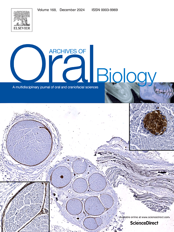
Archives of oral biology
Archives of oral biology 医学-牙科与口腔外科
CiteScore
5.10
自引率
3.30%
发文量
177
审稿时长
26 days
期刊介绍: Archives of Oral Biology is an international journal which aims to publish papers of the highest scientific quality in the oral and craniofacial sciences. The journal is particularly interested in research which advances knowledge in the mechanisms of craniofacial development and disease, including: Cell and molecular biology Molecular genetics Immunology Pathogenesis Cellular microbiology Embryology Syndromology Forensic dentistry
×
引用
GB/T 7714-2015
复制
MLA
复制
APA
复制
导出至
BibTeX EndNote RefMan NoteFirst NoteExpress
×
提示
您的信息不完整,为了账户安全,请先补充。
现在去补充
×
提示
您因"违规操作"
具体请查看互助需知
我知道了
×
提示
确定
请完成安全验证×
copy
已复制链接
快去分享给好友吧!
我知道了
右上角分享
点击右上角分享
0
联系我们:info@booksci.cn Book学术提供免费学术资源搜索服务,方便国内外学者检索中英文文献。致力于提供最便捷和优质的服务体验。 Copyright © 2023 布克学术 All rights reserved.
京ICP备2023020795号-1
ghs 京公网安备 11010802042870号
Book学术文献互助
Book学术文献互助群
群 号:481959085
Book学术官方微信