{"title":"封面图片","authors":"","doi":"10.1002/cm.21957","DOIUrl":null,"url":null,"abstract":"<p>ON THE FRONT COVER: Artistic representation of an expansion microscopy image of human kidney tubular epithelium. Tissues were immunostained for primary cilia (acetylated α-tubulin, yellow), cell-cell junctions (E-cadherin, green/blue), and nuclei (DAPI, dark grey). The cover image is based on the article “Ultrastructure expansion microscopy (U-ExM) of mouse and human kidneys for analysis of subcellular structures”.</p><p>Credit: Ewa Langner and Moe Mahjoub (Department of Medicine, Washington University, St Louis, MO).\n\n <figure>\n <div><picture>\n <source></source></picture><p></p>\n </div>\n </figure>\n </p>","PeriodicalId":55186,"journal":{"name":"Cytoskeleton","volume":"81 11","pages":"C1"},"PeriodicalIF":2.4000,"publicationDate":"2024-11-19","publicationTypes":"Journal Article","fieldsOfStudy":null,"isOpenAccess":false,"openAccessPdf":"https://onlinelibrary.wiley.com/doi/epdf/10.1002/cm.21957","citationCount":"0","resultStr":"{\"title\":\"Front Cover Image\",\"authors\":\"\",\"doi\":\"10.1002/cm.21957\",\"DOIUrl\":null,\"url\":null,\"abstract\":\"<p>ON THE FRONT COVER: Artistic representation of an expansion microscopy image of human kidney tubular epithelium. Tissues were immunostained for primary cilia (acetylated α-tubulin, yellow), cell-cell junctions (E-cadherin, green/blue), and nuclei (DAPI, dark grey). The cover image is based on the article “Ultrastructure expansion microscopy (U-ExM) of mouse and human kidneys for analysis of subcellular structures”.</p><p>Credit: Ewa Langner and Moe Mahjoub (Department of Medicine, Washington University, St Louis, MO).\\n\\n <figure>\\n <div><picture>\\n <source></source></picture><p></p>\\n </div>\\n </figure>\\n </p>\",\"PeriodicalId\":55186,\"journal\":{\"name\":\"Cytoskeleton\",\"volume\":\"81 11\",\"pages\":\"C1\"},\"PeriodicalIF\":2.4000,\"publicationDate\":\"2024-11-19\",\"publicationTypes\":\"Journal Article\",\"fieldsOfStudy\":null,\"isOpenAccess\":false,\"openAccessPdf\":\"https://onlinelibrary.wiley.com/doi/epdf/10.1002/cm.21957\",\"citationCount\":\"0\",\"resultStr\":null,\"platform\":\"Semanticscholar\",\"paperid\":null,\"PeriodicalName\":\"Cytoskeleton\",\"FirstCategoryId\":\"99\",\"ListUrlMain\":\"https://onlinelibrary.wiley.com/doi/10.1002/cm.21957\",\"RegionNum\":4,\"RegionCategory\":\"生物学\",\"ArticlePicture\":[],\"TitleCN\":null,\"AbstractTextCN\":null,\"PMCID\":null,\"EPubDate\":\"\",\"PubModel\":\"\",\"JCR\":\"Q4\",\"JCRName\":\"CELL BIOLOGY\",\"Score\":null,\"Total\":0}","platform":"Semanticscholar","paperid":null,"PeriodicalName":"Cytoskeleton","FirstCategoryId":"99","ListUrlMain":"https://onlinelibrary.wiley.com/doi/10.1002/cm.21957","RegionNum":4,"RegionCategory":"生物学","ArticlePicture":[],"TitleCN":null,"AbstractTextCN":null,"PMCID":null,"EPubDate":"","PubModel":"","JCR":"Q4","JCRName":"CELL BIOLOGY","Score":null,"Total":0}
引用次数: 0
摘要
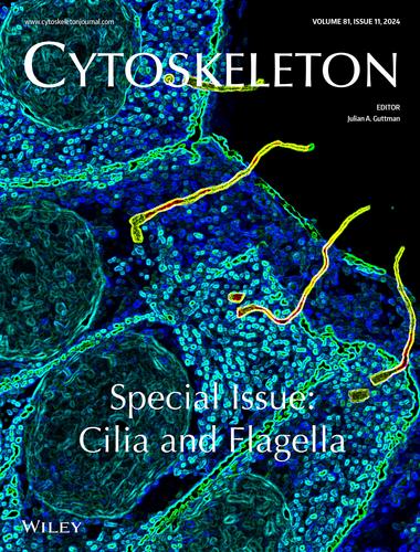
封面:人肾小管上皮细胞扩张显微镜图像的艺术表现。组织上的初级纤毛(乙酰化 α-tubulin,黄色)、细胞-细胞连接(E-cadherin,绿色/蓝色)和细胞核(DAPI,深灰色)均经免疫染色。封面图片来源于文章 "Ultrastructure expansion microscopy (U-ExM) of mouse and human kidneys for analysis of subcellular structures"(用于分析亚细胞结构的小鼠和人类肾脏超微结构扩展显微镜(U-ExM))。
ON THE FRONT COVER: Artistic representation of an expansion microscopy image of human kidney tubular epithelium. Tissues were immunostained for primary cilia (acetylated α-tubulin, yellow), cell-cell junctions (E-cadherin, green/blue), and nuclei (DAPI, dark grey). The cover image is based on the article “Ultrastructure expansion microscopy (U-ExM) of mouse and human kidneys for analysis of subcellular structures”.
Credit: Ewa Langner and Moe Mahjoub (Department of Medicine, Washington University, St Louis, MO).
期刊介绍:
Cytoskeleton focuses on all aspects of cytoskeletal research in healthy and diseased states, spanning genetic and cell biological observations, biochemical, biophysical and structural studies, mathematical modeling and theory. This includes, but is certainly not limited to, classic polymer systems of eukaryotic cells and their structural sites of attachment on membranes and organelles, as well as the bacterial cytoskeleton, the nucleoskeleton, and uncoventional polymer systems with structural/organizational roles. Cytoskeleton is published in 12 issues annually, and special issues will be dedicated to especially-active or newly-emerging areas of cytoskeletal research.