Aman Nikhil, Mudasir Bashir Gugjoo, Ankita Das, Tasaduq Manzoor, Syed Mudasir Ahmad, Nazir Ahmad Ganai, Ashok Kumar
{"title":"富含外泌体的多层低温凝胶可再生并维持山羊骨软骨损伤的软骨结构和表型","authors":"Aman Nikhil, Mudasir Bashir Gugjoo, Ankita Das, Tasaduq Manzoor, Syed Mudasir Ahmad, Nazir Ahmad Ganai, Ashok Kumar","doi":"10.1021/acsami.4c13808","DOIUrl":null,"url":null,"abstract":"<p><p>Treatment of critical-size osteochondral (OC) injuries at load-bearing sites has remained a major clinical challenge in orthopedic surgery. This is due to the anisotropic characteristics of OC tissue and the stratified structure of the cartilage. Here, we developed a multilayered OC scaffold by employing cryogelation technology. Gelatin, chitosan, and chondroitin sulfate were utilized for designing three distinct, 2425 ± 120 μm thick layers of cartilage having different alignments, while nanohydroxyapatite and gelatin were used for the subchondral bone layer. Exosomes derived from articular chondrocytes in the range of 60-110 nm were used to promote chondrogenesis. The biocompatibility and cartilage formation potential of the scaffold and exosomes were initially evaluated in rat OC defects. The application of exosome-loaded scaffolds was then investigated in a critical-size OC injury (8 × 10 mm) created in the goat knee. Artificial synovial fluid was designed and utilized as a carrier for exosomes for a booster dose administered as an intra-articular injection. X-ray imaging and micro-CT analysis revealed that the treatment resulted in improved subchondral bone regeneration. The defect region exhibited healthy hyaline cartilage formation, as detected by MRI imaging. Moreover, histological examination revealed that the treatment group showed augmented cell proliferation, matrix deposition, secretion of proteoglycans, and the formation of stratified hyaline cartilage over a long-term (6 and 12 months), whereas the control group demonstrated the formation of fibrocartilage. Treatment-induced upregulation of collagen II, aggrecan, and SOX 9 genes (∼10 fold) further provided evidence that the cartilage phenotype was well preserved. Hence, the proposed treatment has significant translational potential for treating adverse OC clinical injuries.</p>","PeriodicalId":5,"journal":{"name":"ACS Applied Materials & Interfaces","volume":" ","pages":"64505-64521"},"PeriodicalIF":8.2000,"publicationDate":"2024-11-27","publicationTypes":"Journal Article","fieldsOfStudy":null,"isOpenAccess":false,"openAccessPdf":"","citationCount":"0","resultStr":"{\"title\":\"Multilayered Cryogel Enriched with Exosomes Regenerates and Maintains Cartilage Architecture and Phenotype in Goat Osteochondral Injuries.\",\"authors\":\"Aman Nikhil, Mudasir Bashir Gugjoo, Ankita Das, Tasaduq Manzoor, Syed Mudasir Ahmad, Nazir Ahmad Ganai, Ashok Kumar\",\"doi\":\"10.1021/acsami.4c13808\",\"DOIUrl\":null,\"url\":null,\"abstract\":\"<p><p>Treatment of critical-size osteochondral (OC) injuries at load-bearing sites has remained a major clinical challenge in orthopedic surgery. This is due to the anisotropic characteristics of OC tissue and the stratified structure of the cartilage. Here, we developed a multilayered OC scaffold by employing cryogelation technology. Gelatin, chitosan, and chondroitin sulfate were utilized for designing three distinct, 2425 ± 120 μm thick layers of cartilage having different alignments, while nanohydroxyapatite and gelatin were used for the subchondral bone layer. Exosomes derived from articular chondrocytes in the range of 60-110 nm were used to promote chondrogenesis. The biocompatibility and cartilage formation potential of the scaffold and exosomes were initially evaluated in rat OC defects. The application of exosome-loaded scaffolds was then investigated in a critical-size OC injury (8 × 10 mm) created in the goat knee. Artificial synovial fluid was designed and utilized as a carrier for exosomes for a booster dose administered as an intra-articular injection. X-ray imaging and micro-CT analysis revealed that the treatment resulted in improved subchondral bone regeneration. The defect region exhibited healthy hyaline cartilage formation, as detected by MRI imaging. Moreover, histological examination revealed that the treatment group showed augmented cell proliferation, matrix deposition, secretion of proteoglycans, and the formation of stratified hyaline cartilage over a long-term (6 and 12 months), whereas the control group demonstrated the formation of fibrocartilage. Treatment-induced upregulation of collagen II, aggrecan, and SOX 9 genes (∼10 fold) further provided evidence that the cartilage phenotype was well preserved. Hence, the proposed treatment has significant translational potential for treating adverse OC clinical injuries.</p>\",\"PeriodicalId\":5,\"journal\":{\"name\":\"ACS Applied Materials & Interfaces\",\"volume\":\" \",\"pages\":\"64505-64521\"},\"PeriodicalIF\":8.2000,\"publicationDate\":\"2024-11-27\",\"publicationTypes\":\"Journal Article\",\"fieldsOfStudy\":null,\"isOpenAccess\":false,\"openAccessPdf\":\"\",\"citationCount\":\"0\",\"resultStr\":null,\"platform\":\"Semanticscholar\",\"paperid\":null,\"PeriodicalName\":\"ACS Applied Materials & Interfaces\",\"FirstCategoryId\":\"88\",\"ListUrlMain\":\"https://doi.org/10.1021/acsami.4c13808\",\"RegionNum\":2,\"RegionCategory\":\"材料科学\",\"ArticlePicture\":[],\"TitleCN\":null,\"AbstractTextCN\":null,\"PMCID\":null,\"EPubDate\":\"2024/11/18 0:00:00\",\"PubModel\":\"Epub\",\"JCR\":\"Q1\",\"JCRName\":\"MATERIALS SCIENCE, MULTIDISCIPLINARY\",\"Score\":null,\"Total\":0}","platform":"Semanticscholar","paperid":null,"PeriodicalName":"ACS Applied Materials & Interfaces","FirstCategoryId":"88","ListUrlMain":"https://doi.org/10.1021/acsami.4c13808","RegionNum":2,"RegionCategory":"材料科学","ArticlePicture":[],"TitleCN":null,"AbstractTextCN":null,"PMCID":null,"EPubDate":"2024/11/18 0:00:00","PubModel":"Epub","JCR":"Q1","JCRName":"MATERIALS SCIENCE, MULTIDISCIPLINARY","Score":null,"Total":0}
Multilayered Cryogel Enriched with Exosomes Regenerates and Maintains Cartilage Architecture and Phenotype in Goat Osteochondral Injuries.
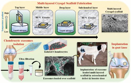
Treatment of critical-size osteochondral (OC) injuries at load-bearing sites has remained a major clinical challenge in orthopedic surgery. This is due to the anisotropic characteristics of OC tissue and the stratified structure of the cartilage. Here, we developed a multilayered OC scaffold by employing cryogelation technology. Gelatin, chitosan, and chondroitin sulfate were utilized for designing three distinct, 2425 ± 120 μm thick layers of cartilage having different alignments, while nanohydroxyapatite and gelatin were used for the subchondral bone layer. Exosomes derived from articular chondrocytes in the range of 60-110 nm were used to promote chondrogenesis. The biocompatibility and cartilage formation potential of the scaffold and exosomes were initially evaluated in rat OC defects. The application of exosome-loaded scaffolds was then investigated in a critical-size OC injury (8 × 10 mm) created in the goat knee. Artificial synovial fluid was designed and utilized as a carrier for exosomes for a booster dose administered as an intra-articular injection. X-ray imaging and micro-CT analysis revealed that the treatment resulted in improved subchondral bone regeneration. The defect region exhibited healthy hyaline cartilage formation, as detected by MRI imaging. Moreover, histological examination revealed that the treatment group showed augmented cell proliferation, matrix deposition, secretion of proteoglycans, and the formation of stratified hyaline cartilage over a long-term (6 and 12 months), whereas the control group demonstrated the formation of fibrocartilage. Treatment-induced upregulation of collagen II, aggrecan, and SOX 9 genes (∼10 fold) further provided evidence that the cartilage phenotype was well preserved. Hence, the proposed treatment has significant translational potential for treating adverse OC clinical injuries.
期刊介绍:
ACS Applied Materials & Interfaces is a leading interdisciplinary journal that brings together chemists, engineers, physicists, and biologists to explore the development and utilization of newly-discovered materials and interfacial processes for specific applications. Our journal has experienced remarkable growth since its establishment in 2009, both in terms of the number of articles published and the impact of the research showcased. We are proud to foster a truly global community, with the majority of published articles originating from outside the United States, reflecting the rapid growth of applied research worldwide.
 求助内容:
求助内容: 应助结果提醒方式:


