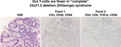
在 "完全 "22q11.2缺失综合征(迪乔治综合征)患者的胃肠道管腔中,T细胞明显减少:利用色原多重免疫组化技术确定细胞群。

IF 1.5 4区 医学 Q3 PATHOLOGY
Grace J. Kwon , William R. Jeck , Shannon McCall , Zuowei Su , Avani A. Pendse
{"title":"在 \"完全 \"22q11.2缺失综合征(迪乔治综合征)患者的胃肠道管腔中,T细胞明显减少:利用色原多重免疫组化技术确定细胞群。","authors":"Grace J. Kwon ,&nbsp;William R. Jeck ,&nbsp;Shannon McCall ,&nbsp;Zuowei Su ,&nbsp;Avani A. Pendse","doi":"10.1016/j.anndiagpath.2024.152391","DOIUrl":null,"url":null,"abstract":"<div><div>Patients with 22q11.2 deletion syndrome or DiGeorge syndrome commonly report gastrointestinal symptoms in addition to more widely understood cardiac and immunodeficiency abnormalities. However, the morphologic features of gastrointestinal tract pathology in these patients are poorly understood. We previously reported that plasma cells are essentially absent from the luminal gastrointestinal tract of patients with “complete” DiGeorge syndrome. Herein, we add to the current understanding of the luminal gastrointestinal tract changes in patients with DiGeorge syndrome. Patients with cytogenetically confirmed DiGeorge syndrome were identified after approval from our institutional review board. Gastrointestinal tract biopsies from patients with DiGeorge syndrome that were severely immunosuppressed (complete DiGeorge syndrome, DGS-I), partially immunocompromised (partial DiGeorge syndrome, DGS), and from control patients were reviewed. Two panels of chromogenic multiplex immunohistochemistry (IHC) were performed to evaluate the immune cell infiltrate in the lamina propria of the duodenum and colon. “Panel #1” was composed of antibodies targeting CD3, CD20, and CD68. “Panel #2” was composed of antibodies targeting CD4, CD8, CD56, and TCRϒδ. Assessment of cell types identified by these antibody targets demonstrated a significant reduction of duodenal and colonic T-cells in patients with complete DiGeorge syndrome. In addition to establishing the morphologic phenotype of the luminal gastrointestinal tract of patients with DiGeorge syndrome, we also highlight our chosen technology of chromogenic multiplex IHC as a relatively accessible research and diagnostic tool with wide potential to be utilized across various disease processes.</div></div>","PeriodicalId":50768,"journal":{"name":"Annals of Diagnostic Pathology","volume":"74 ","pages":"Article 152391"},"PeriodicalIF":1.5000,"publicationDate":"2024-11-06","publicationTypes":"Journal Article","fieldsOfStudy":null,"isOpenAccess":false,"openAccessPdf":"","citationCount":"0","resultStr":"{\"title\":\"T-cells are significantly reduced in the luminal gastrointestinal tract of patients with “complete” 22q11.2 deletion syndrome (DiGeorge syndrome): Utilization of chromogenic multiplex immunohistochemistry to define cellular populations\",\"authors\":\"Grace J. Kwon ,&nbsp;William R. Jeck ,&nbsp;Shannon McCall ,&nbsp;Zuowei Su ,&nbsp;Avani A. Pendse\",\"doi\":\"10.1016/j.anndiagpath.2024.152391\",\"DOIUrl\":null,\"url\":null,\"abstract\":\"<div><div>Patients with 22q11.2 deletion syndrome or DiGeorge syndrome commonly report gastrointestinal symptoms in addition to more widely understood cardiac and immunodeficiency abnormalities. However, the morphologic features of gastrointestinal tract pathology in these patients are poorly understood. We previously reported that plasma cells are essentially absent from the luminal gastrointestinal tract of patients with “complete” DiGeorge syndrome. Herein, we add to the current understanding of the luminal gastrointestinal tract changes in patients with DiGeorge syndrome. Patients with cytogenetically confirmed DiGeorge syndrome were identified after approval from our institutional review board. Gastrointestinal tract biopsies from patients with DiGeorge syndrome that were severely immunosuppressed (complete DiGeorge syndrome, DGS-I), partially immunocompromised (partial DiGeorge syndrome, DGS), and from control patients were reviewed. Two panels of chromogenic multiplex immunohistochemistry (IHC) were performed to evaluate the immune cell infiltrate in the lamina propria of the duodenum and colon. “Panel #1” was composed of antibodies targeting CD3, CD20, and CD68. “Panel #2” was composed of antibodies targeting CD4, CD8, CD56, and TCRϒδ. Assessment of cell types identified by these antibody targets demonstrated a significant reduction of duodenal and colonic T-cells in patients with complete DiGeorge syndrome. In addition to establishing the morphologic phenotype of the luminal gastrointestinal tract of patients with DiGeorge syndrome, we also highlight our chosen technology of chromogenic multiplex IHC as a relatively accessible research and diagnostic tool with wide potential to be utilized across various disease processes.</div></div>\",\"PeriodicalId\":50768,\"journal\":{\"name\":\"Annals of Diagnostic Pathology\",\"volume\":\"74 \",\"pages\":\"Article 152391\"},\"PeriodicalIF\":1.5000,\"publicationDate\":\"2024-11-06\",\"publicationTypes\":\"Journal Article\",\"fieldsOfStudy\":null,\"isOpenAccess\":false,\"openAccessPdf\":\"\",\"citationCount\":\"0\",\"resultStr\":null,\"platform\":\"Semanticscholar\",\"paperid\":null,\"PeriodicalName\":\"Annals of Diagnostic Pathology\",\"FirstCategoryId\":\"3\",\"ListUrlMain\":\"https://www.sciencedirect.com/science/article/pii/S109291342400128X\",\"RegionNum\":4,\"RegionCategory\":\"医学\",\"ArticlePicture\":[],\"TitleCN\":null,\"AbstractTextCN\":null,\"PMCID\":null,\"EPubDate\":\"\",\"PubModel\":\"\",\"JCR\":\"Q3\",\"JCRName\":\"PATHOLOGY\",\"Score\":null,\"Total\":0}","platform":"Semanticscholar","paperid":null,"PeriodicalName":"Annals of Diagnostic Pathology","FirstCategoryId":"3","ListUrlMain":"https://www.sciencedirect.com/science/article/pii/S109291342400128X","RegionNum":4,"RegionCategory":"医学","ArticlePicture":[],"TitleCN":null,"AbstractTextCN":null,"PMCID":null,"EPubDate":"","PubModel":"","JCR":"Q3","JCRName":"PATHOLOGY","Score":null,"Total":0}
引用次数: 0

摘要

22q11.2缺失综合征或迪乔治综合征患者除了普遍存在的心脏和免疫缺陷异常外,通常还伴有胃肠道症状。然而,人们对这些患者胃肠道病变的形态特征却知之甚少。我们以前曾报道,"完全型 "迪乔治综合征患者的胃肠道管腔内基本上没有浆细胞。在此,我们将进一步阐述目前对迪乔治综合征患者胃肠道腔内变化的认识。经本机构审查委员会批准后,我们确定了经细胞遗传学确诊的迪乔治综合征患者。对免疫抑制严重的迪乔治综合征患者(完全性迪乔治综合征,DGS-I)、免疫功能部分低下的患者(部分性迪乔治综合征,DGS)以及对照组患者的胃肠道活检组织进行了审查。为评估十二指肠和结肠固有层的免疫细胞浸润情况,采用了两组色原多重免疫组化(IHC)。"面板 #1 "由针对 CD3、CD20 和 CD68 的抗体组成。"面板 2 "由针对 CD4、CD8、CD56 和 TCRϒδ 的抗体组成。通过评估这些抗体靶标确定的细胞类型,发现完全性迪乔治综合征患者的十二指肠和结肠 T 细胞明显减少。除了确定迪乔治综合征患者管腔胃肠道的形态表型外,我们还强调了我们所选择的色原多重 IHC 技术,它是一种相对容易获得的研究和诊断工具,在各种疾病过程中具有广泛的应用潜力。
本文章由计算机程序翻译,如有差异,请以英文原文为准。

T-cells are significantly reduced in the luminal gastrointestinal tract of patients with “complete” 22q11.2 deletion syndrome (DiGeorge syndrome): Utilization of chromogenic multiplex immunohistochemistry to define cellular populations

T-cells are significantly reduced in the luminal gastrointestinal tract of patients with “complete” 22q11.2 deletion syndrome (DiGeorge syndrome): Utilization of chromogenic multiplex immunohistochemistry to define cellular populations
Patients with 22q11.2 deletion syndrome or DiGeorge syndrome commonly report gastrointestinal symptoms in addition to more widely understood cardiac and immunodeficiency abnormalities. However, the morphologic features of gastrointestinal tract pathology in these patients are poorly understood. We previously reported that plasma cells are essentially absent from the luminal gastrointestinal tract of patients with “complete” DiGeorge syndrome. Herein, we add to the current understanding of the luminal gastrointestinal tract changes in patients with DiGeorge syndrome. Patients with cytogenetically confirmed DiGeorge syndrome were identified after approval from our institutional review board. Gastrointestinal tract biopsies from patients with DiGeorge syndrome that were severely immunosuppressed (complete DiGeorge syndrome, DGS-I), partially immunocompromised (partial DiGeorge syndrome, DGS), and from control patients were reviewed. Two panels of chromogenic multiplex immunohistochemistry (IHC) were performed to evaluate the immune cell infiltrate in the lamina propria of the duodenum and colon. “Panel #1” was composed of antibodies targeting CD3, CD20, and CD68. “Panel #2” was composed of antibodies targeting CD4, CD8, CD56, and TCRϒδ. Assessment of cell types identified by these antibody targets demonstrated a significant reduction of duodenal and colonic T-cells in patients with complete DiGeorge syndrome. In addition to establishing the morphologic phenotype of the luminal gastrointestinal tract of patients with DiGeorge syndrome, we also highlight our chosen technology of chromogenic multiplex IHC as a relatively accessible research and diagnostic tool with wide potential to be utilized across various disease processes.
求助全文
通过发布文献求助,成功后即可免费获取论文全文。 去求助
来源期刊
CiteScore
3.90
自引率
5.00%
发文量
149
审稿时长
26 days
期刊介绍: A peer-reviewed journal devoted to the publication of articles dealing with traditional morphologic studies using standard diagnostic techniques and stressing clinicopathological correlations and scientific observation of relevance to the daily practice of pathology. Special features include pathologic-radiologic correlations and pathologic-cytologic correlations.
×
引用
GB/T 7714-2015
复制
MLA
复制
APA
复制
导出至
BibTeX EndNote RefMan NoteFirst NoteExpress
×
提示
您的信息不完整,为了账户安全,请先补充。
现在去补充
×
提示
您因"违规操作"
具体请查看互助需知
我知道了
×
提示
确定
请完成安全验证×
copy
已复制链接
快去分享给好友吧!
我知道了
右上角分享
点击右上角分享
0
联系我们:info@booksci.cn Book学术提供免费学术资源搜索服务,方便国内外学者检索中英文文献。致力于提供最便捷和优质的服务体验。 Copyright © 2023 布克学术 All rights reserved.
京ICP备2023020795号-1
ghs 京公网安备 11010802042870号
Book学术文献互助
Book学术文献互助群
群 号:481959085
Book学术官方微信