Nicole Wake, Yenpo Lin, Ek T Tan, Darryl B Sneag, Sarah Ianucci, Maggie Fung
{"title":"利用磁共振神经成像技术三维打印臂丛及其骨性地标,用于胸廓出口综合征评估。","authors":"Nicole Wake, Yenpo Lin, Ek T Tan, Darryl B Sneag, Sarah Ianucci, Maggie Fung","doi":"10.1186/s41205-024-00239-6","DOIUrl":null,"url":null,"abstract":"<p><strong>Background: </strong>Patient-specific three-dimensional (3D) printed anatomic models are valuable clinical tools that facilitate enhanced visualization of pertinent anatomic structures and have demonstrated benefits of reduced surgical times, increased surgeon confidence, and improved operative results and subsequent patient outcomes. Medical image-based 3D printed anatomic models are generally created from computed tomography (CT), however magnetic resonance imaging (MRI), which offers exquisite soft tissue characterization and flexible contrast avoiding the use of ionizing radiation, is an attractive alternative. Herein, the application of 3D printing incorporating both MR neurography and zero-echo time (ZTE) MRI for visualization of the brachial plexus anatomy in a subject with thoracic outlet syndrome (TOS) is described.</p><p><strong>Methods: </strong>A 28-year-old man presented with chronic right upper limb discomfort and paresthesias extending from the shoulder region to the third and fourth digits. The subject underwent evaluation with a unilateral brachial plexus MR neurography protocol at 3.0 Tesla for suspicion of TOS. The protocol included T2-weighted, 3D fast spin echo short-tau inversion recovery (STIR-FSE) and 3D radial ZTE sequences for depiction of the nerves and bones, respectively. The first rib and its synostosis impinged upon the inferior aspect of the T1 nerve root (T1NR), with accompanying mild enlargement of the T1NR. A 3D printed anatomic model was created and included: (1) bone (spine, ribs, clavicle, scapula, and humerus), (2) brachial plexus, and (3) costal cartilage.</p><p><strong>Results: </strong>The 3D printed model clearly demonstrated a T1NR impingement from the synostosis, confirming the diagnosis of neurologic thoracic outlet syndrome (TOS) and guided the treatment approach in prescribing TOS-specific physical therapy, which led to significant improvements in the patient's condition.</p><p><strong>Conclusion: </strong>To our knowledge, this is the first in-vivo human 3D printed case for TOS using MRI-only data. The 3D printed model allowed for improved visualization and understanding of the spatial relationships between the nerves of the brachial plexus and surrounding osseous structures responsible for the patient's symptoms.</p><p><strong>Clinical trial number: </strong>Not applicable.</p>","PeriodicalId":72036,"journal":{"name":"3D printing in medicine","volume":"10 1","pages":"36"},"PeriodicalIF":3.1000,"publicationDate":"2024-11-14","publicationTypes":"Journal Article","fieldsOfStudy":null,"isOpenAccess":false,"openAccessPdf":"https://www.ncbi.nlm.nih.gov/pmc/articles/PMC11562346/pdf/","citationCount":"0","resultStr":"{\"title\":\"3D printing of the brachial plexus and its osseous landmarks using magnetic resonance neurography for thoracic outlet syndrome evaluation.\",\"authors\":\"Nicole Wake, Yenpo Lin, Ek T Tan, Darryl B Sneag, Sarah Ianucci, Maggie Fung\",\"doi\":\"10.1186/s41205-024-00239-6\",\"DOIUrl\":null,\"url\":null,\"abstract\":\"<p><strong>Background: </strong>Patient-specific three-dimensional (3D) printed anatomic models are valuable clinical tools that facilitate enhanced visualization of pertinent anatomic structures and have demonstrated benefits of reduced surgical times, increased surgeon confidence, and improved operative results and subsequent patient outcomes. Medical image-based 3D printed anatomic models are generally created from computed tomography (CT), however magnetic resonance imaging (MRI), which offers exquisite soft tissue characterization and flexible contrast avoiding the use of ionizing radiation, is an attractive alternative. Herein, the application of 3D printing incorporating both MR neurography and zero-echo time (ZTE) MRI for visualization of the brachial plexus anatomy in a subject with thoracic outlet syndrome (TOS) is described.</p><p><strong>Methods: </strong>A 28-year-old man presented with chronic right upper limb discomfort and paresthesias extending from the shoulder region to the third and fourth digits. The subject underwent evaluation with a unilateral brachial plexus MR neurography protocol at 3.0 Tesla for suspicion of TOS. The protocol included T2-weighted, 3D fast spin echo short-tau inversion recovery (STIR-FSE) and 3D radial ZTE sequences for depiction of the nerves and bones, respectively. The first rib and its synostosis impinged upon the inferior aspect of the T1 nerve root (T1NR), with accompanying mild enlargement of the T1NR. A 3D printed anatomic model was created and included: (1) bone (spine, ribs, clavicle, scapula, and humerus), (2) brachial plexus, and (3) costal cartilage.</p><p><strong>Results: </strong>The 3D printed model clearly demonstrated a T1NR impingement from the synostosis, confirming the diagnosis of neurologic thoracic outlet syndrome (TOS) and guided the treatment approach in prescribing TOS-specific physical therapy, which led to significant improvements in the patient's condition.</p><p><strong>Conclusion: </strong>To our knowledge, this is the first in-vivo human 3D printed case for TOS using MRI-only data. The 3D printed model allowed for improved visualization and understanding of the spatial relationships between the nerves of the brachial plexus and surrounding osseous structures responsible for the patient's symptoms.</p><p><strong>Clinical trial number: </strong>Not applicable.</p>\",\"PeriodicalId\":72036,\"journal\":{\"name\":\"3D printing in medicine\",\"volume\":\"10 1\",\"pages\":\"36\"},\"PeriodicalIF\":3.1000,\"publicationDate\":\"2024-11-14\",\"publicationTypes\":\"Journal Article\",\"fieldsOfStudy\":null,\"isOpenAccess\":false,\"openAccessPdf\":\"https://www.ncbi.nlm.nih.gov/pmc/articles/PMC11562346/pdf/\",\"citationCount\":\"0\",\"resultStr\":null,\"platform\":\"Semanticscholar\",\"paperid\":null,\"PeriodicalName\":\"3D printing in medicine\",\"FirstCategoryId\":\"1085\",\"ListUrlMain\":\"https://doi.org/10.1186/s41205-024-00239-6\",\"RegionNum\":0,\"RegionCategory\":null,\"ArticlePicture\":[],\"TitleCN\":null,\"AbstractTextCN\":null,\"PMCID\":null,\"EPubDate\":\"\",\"PubModel\":\"\",\"JCR\":\"Q1\",\"JCRName\":\"RADIOLOGY, NUCLEAR MEDICINE & MEDICAL IMAGING\",\"Score\":null,\"Total\":0}","platform":"Semanticscholar","paperid":null,"PeriodicalName":"3D printing in medicine","FirstCategoryId":"1085","ListUrlMain":"https://doi.org/10.1186/s41205-024-00239-6","RegionNum":0,"RegionCategory":null,"ArticlePicture":[],"TitleCN":null,"AbstractTextCN":null,"PMCID":null,"EPubDate":"","PubModel":"","JCR":"Q1","JCRName":"RADIOLOGY, NUCLEAR MEDICINE & MEDICAL IMAGING","Score":null,"Total":0}
引用次数: 0
摘要
背景:患者特异性三维(3D)打印解剖模型是非常有价值的临床工具,有助于增强相关解剖结构的可视化,并具有缩短手术时间、增强外科医生信心、改善手术效果和患者后续预后等优点。基于医学影像的 3D 打印解剖模型通常是通过计算机断层扫描(CT)创建的,但磁共振成像(MRI)可提供细腻的软组织特征和灵活的对比度,避免使用电离辐射,是一种极具吸引力的替代方法。本文介绍了结合磁共振神经成像和零回波时间(ZTE)磁共振成像的 3D 打印技术在胸廓出口综合征(TOS)患者臂丛神经解剖学可视化中的应用:方法:一名 28 岁的男子因慢性右上肢不适和从肩部延伸至第三和第四个手指的麻痹而就诊。该患者因怀疑患有 TOS 而接受了 3.0 特斯拉单侧臂丛磁共振神经成像检查。该方案包括 T2 加权、三维快速自旋回波短陶反转恢复(STIR-FSE)和三维径向 ZTE 序列,分别用于描绘神经和骨骼。第一根肋骨及其突起物侵犯了 T1 神经根(T1NR)的下侧,并伴有 T1NR 的轻度增大。创建的 3D 打印解剖模型包括(结果:结果:3D 打印模型清楚地显示了来自关节突的 T1NR 撞击,证实了神经性胸廓出口综合征(TOS)的诊断,并指导了治疗方法,为患者开出了针对 TOS 的物理治疗处方,使患者的病情得到了显著改善:据我们所知,这是首个使用纯核磁共振成像数据的TOS体内人体3D打印病例。三维打印模型提高了对臂丛神经和导致患者症状的周围骨性结构之间空间关系的可视化和理解:临床试验编号:不适用。
3D printing of the brachial plexus and its osseous landmarks using magnetic resonance neurography for thoracic outlet syndrome evaluation.
Background: Patient-specific three-dimensional (3D) printed anatomic models are valuable clinical tools that facilitate enhanced visualization of pertinent anatomic structures and have demonstrated benefits of reduced surgical times, increased surgeon confidence, and improved operative results and subsequent patient outcomes. Medical image-based 3D printed anatomic models are generally created from computed tomography (CT), however magnetic resonance imaging (MRI), which offers exquisite soft tissue characterization and flexible contrast avoiding the use of ionizing radiation, is an attractive alternative. Herein, the application of 3D printing incorporating both MR neurography and zero-echo time (ZTE) MRI for visualization of the brachial plexus anatomy in a subject with thoracic outlet syndrome (TOS) is described.
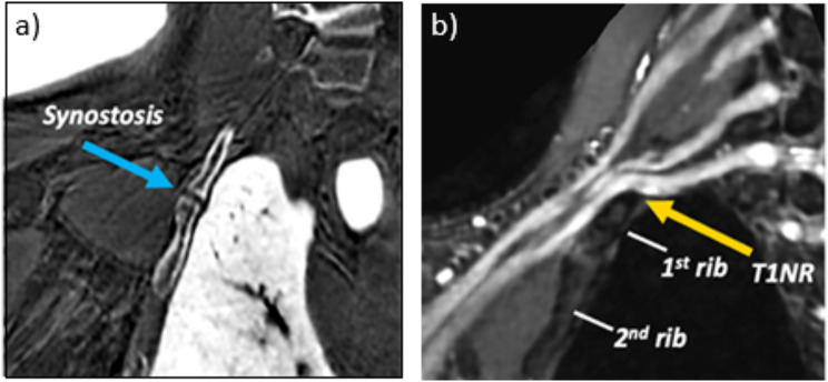
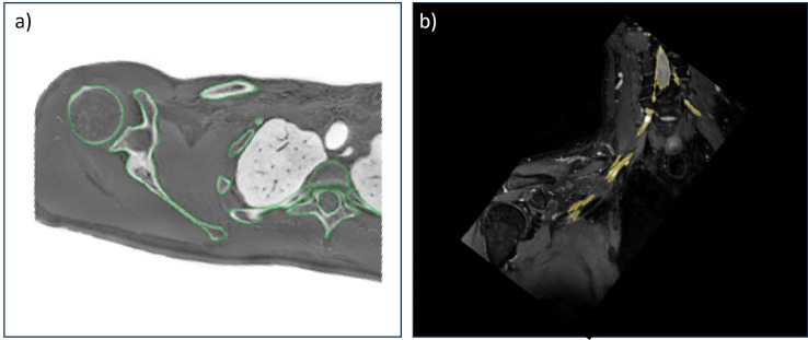
Methods: A 28-year-old man presented with chronic right upper limb discomfort and paresthesias extending from the shoulder region to the third and fourth digits. The subject underwent evaluation with a unilateral brachial plexus MR neurography protocol at 3.0 Tesla for suspicion of TOS. The protocol included T2-weighted, 3D fast spin echo short-tau inversion recovery (STIR-FSE) and 3D radial ZTE sequences for depiction of the nerves and bones, respectively. The first rib and its synostosis impinged upon the inferior aspect of the T1 nerve root (T1NR), with accompanying mild enlargement of the T1NR. A 3D printed anatomic model was created and included: (1) bone (spine, ribs, clavicle, scapula, and humerus), (2) brachial plexus, and (3) costal cartilage.
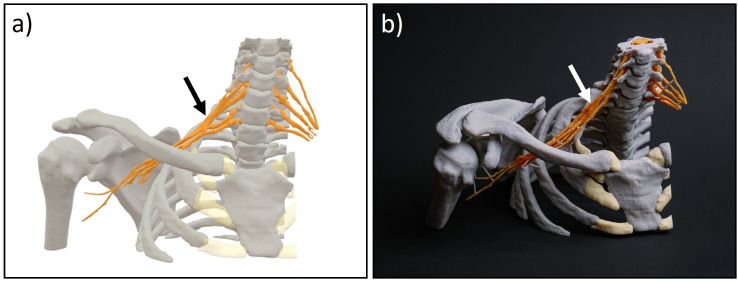
Results: The 3D printed model clearly demonstrated a T1NR impingement from the synostosis, confirming the diagnosis of neurologic thoracic outlet syndrome (TOS) and guided the treatment approach in prescribing TOS-specific physical therapy, which led to significant improvements in the patient's condition.
Conclusion: To our knowledge, this is the first in-vivo human 3D printed case for TOS using MRI-only data. The 3D printed model allowed for improved visualization and understanding of the spatial relationships between the nerves of the brachial plexus and surrounding osseous structures responsible for the patient's symptoms.

 求助内容:
求助内容: 应助结果提醒方式:


