Daohuan Lu , Kehan Cai , Zhiwen Zeng , Jun Huang , Nianfang Ma , Botao Gao , Shan Yu
{"title":"载入血管内皮生长因子的肝素化透明质酸大孔水凝胶,用于增强三维内皮细胞迁移和血管形成。","authors":"Daohuan Lu , Kehan Cai , Zhiwen Zeng , Jun Huang , Nianfang Ma , Botao Gao , Shan Yu","doi":"10.1016/j.bioadv.2024.214094","DOIUrl":null,"url":null,"abstract":"<div><div>The formation of robust vascular systems within voluminous scaffolds remains a formidable barrier in the realm of tissue engineering. There is a growing interest in the integration of biomaterial scaffolds with multiple physical and chemical stimuli to augment the process of vascularization. This study aims to investigate the combined impact of macroporous structures and vascular endothelial growth factor (VEGF) on cell migration and vascularization. Heparinized hyaluronic acid (HepHA) macroporous hydrogels with differing pore sizes, composed by methacrylated hyaluronic acid (HAMA) and methacrylated heparin (HepMA), were fabricated by a gelatin microspheres (GMS) template leaching method. After characterization of their physical properties, VEGF was immobilized on the HepHA hydrogels. The <em>in vitro</em> release study indicated that the HepHA hydrogels can provide sustained release of VEGF. Subsequently, cells migration of human umbilical vein endothelial (HUVECs) assessment indicated that HUVECs cultured on VEGF-loaded HepHA hydrogels with larger pores (VEGF@HepHA250) migrated the furthest. Finally, the hydrogels were implanted and evaluated using a dorsal subcutaneous model. The histological analyses conducted <em>in vivo</em> were consistent with the <em>in vitro</em> results, VEGF@HepHA250 hydrogels exhibited the most pronounced vascularization four weeks post-implantation, indicating that hydrogels with expanded pores and an enriched VEGF promoted angiogenesis within the hydrogels. This study sheds light on the synergistic effects of VEGF release on 3D cell migration and vascularization within hydrogels of differing pore sizes, thus providing novel insights into the strategic design and fabrication of tissue-engineered scaffolds that are amenable to vascularization.</div></div>","PeriodicalId":51111,"journal":{"name":"Materials Science & Engineering C-Materials for Biological Applications","volume":"167 ","pages":"Article 214094"},"PeriodicalIF":5.5000,"publicationDate":"2024-10-31","publicationTypes":"Journal Article","fieldsOfStudy":null,"isOpenAccess":false,"openAccessPdf":"","citationCount":"0","resultStr":"{\"title\":\"VEGF loading heparinized hyaluronic acid macroporous hydrogels for enhanced 3D endothelial cell migration and vascularization\",\"authors\":\"Daohuan Lu , Kehan Cai , Zhiwen Zeng , Jun Huang , Nianfang Ma , Botao Gao , Shan Yu\",\"doi\":\"10.1016/j.bioadv.2024.214094\",\"DOIUrl\":null,\"url\":null,\"abstract\":\"<div><div>The formation of robust vascular systems within voluminous scaffolds remains a formidable barrier in the realm of tissue engineering. There is a growing interest in the integration of biomaterial scaffolds with multiple physical and chemical stimuli to augment the process of vascularization. This study aims to investigate the combined impact of macroporous structures and vascular endothelial growth factor (VEGF) on cell migration and vascularization. Heparinized hyaluronic acid (HepHA) macroporous hydrogels with differing pore sizes, composed by methacrylated hyaluronic acid (HAMA) and methacrylated heparin (HepMA), were fabricated by a gelatin microspheres (GMS) template leaching method. After characterization of their physical properties, VEGF was immobilized on the HepHA hydrogels. The <em>in vitro</em> release study indicated that the HepHA hydrogels can provide sustained release of VEGF. Subsequently, cells migration of human umbilical vein endothelial (HUVECs) assessment indicated that HUVECs cultured on VEGF-loaded HepHA hydrogels with larger pores (VEGF@HepHA250) migrated the furthest. Finally, the hydrogels were implanted and evaluated using a dorsal subcutaneous model. The histological analyses conducted <em>in vivo</em> were consistent with the <em>in vitro</em> results, VEGF@HepHA250 hydrogels exhibited the most pronounced vascularization four weeks post-implantation, indicating that hydrogels with expanded pores and an enriched VEGF promoted angiogenesis within the hydrogels. This study sheds light on the synergistic effects of VEGF release on 3D cell migration and vascularization within hydrogels of differing pore sizes, thus providing novel insights into the strategic design and fabrication of tissue-engineered scaffolds that are amenable to vascularization.</div></div>\",\"PeriodicalId\":51111,\"journal\":{\"name\":\"Materials Science & Engineering C-Materials for Biological Applications\",\"volume\":\"167 \",\"pages\":\"Article 214094\"},\"PeriodicalIF\":5.5000,\"publicationDate\":\"2024-10-31\",\"publicationTypes\":\"Journal Article\",\"fieldsOfStudy\":null,\"isOpenAccess\":false,\"openAccessPdf\":\"\",\"citationCount\":\"0\",\"resultStr\":null,\"platform\":\"Semanticscholar\",\"paperid\":null,\"PeriodicalName\":\"Materials Science & Engineering C-Materials for Biological Applications\",\"FirstCategoryId\":\"1085\",\"ListUrlMain\":\"https://www.sciencedirect.com/science/article/pii/S2772950824003376\",\"RegionNum\":2,\"RegionCategory\":\"医学\",\"ArticlePicture\":[],\"TitleCN\":null,\"AbstractTextCN\":null,\"PMCID\":null,\"EPubDate\":\"\",\"PubModel\":\"\",\"JCR\":\"Q2\",\"JCRName\":\"MATERIALS SCIENCE, BIOMATERIALS\",\"Score\":null,\"Total\":0}","platform":"Semanticscholar","paperid":null,"PeriodicalName":"Materials Science & Engineering C-Materials for Biological Applications","FirstCategoryId":"1085","ListUrlMain":"https://www.sciencedirect.com/science/article/pii/S2772950824003376","RegionNum":2,"RegionCategory":"医学","ArticlePicture":[],"TitleCN":null,"AbstractTextCN":null,"PMCID":null,"EPubDate":"","PubModel":"","JCR":"Q2","JCRName":"MATERIALS SCIENCE, BIOMATERIALS","Score":null,"Total":0}
VEGF loading heparinized hyaluronic acid macroporous hydrogels for enhanced 3D endothelial cell migration and vascularization
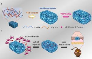
The formation of robust vascular systems within voluminous scaffolds remains a formidable barrier in the realm of tissue engineering. There is a growing interest in the integration of biomaterial scaffolds with multiple physical and chemical stimuli to augment the process of vascularization. This study aims to investigate the combined impact of macroporous structures and vascular endothelial growth factor (VEGF) on cell migration and vascularization. Heparinized hyaluronic acid (HepHA) macroporous hydrogels with differing pore sizes, composed by methacrylated hyaluronic acid (HAMA) and methacrylated heparin (HepMA), were fabricated by a gelatin microspheres (GMS) template leaching method. After characterization of their physical properties, VEGF was immobilized on the HepHA hydrogels. The in vitro release study indicated that the HepHA hydrogels can provide sustained release of VEGF. Subsequently, cells migration of human umbilical vein endothelial (HUVECs) assessment indicated that HUVECs cultured on VEGF-loaded HepHA hydrogels with larger pores (VEGF@HepHA250) migrated the furthest. Finally, the hydrogels were implanted and evaluated using a dorsal subcutaneous model. The histological analyses conducted in vivo were consistent with the in vitro results, VEGF@HepHA250 hydrogels exhibited the most pronounced vascularization four weeks post-implantation, indicating that hydrogels with expanded pores and an enriched VEGF promoted angiogenesis within the hydrogels. This study sheds light on the synergistic effects of VEGF release on 3D cell migration and vascularization within hydrogels of differing pore sizes, thus providing novel insights into the strategic design and fabrication of tissue-engineered scaffolds that are amenable to vascularization.
期刊介绍:
Biomaterials Advances, previously known as Materials Science and Engineering: C-Materials for Biological Applications (P-ISSN: 0928-4931, E-ISSN: 1873-0191). Includes topics at the interface of the biomedical sciences and materials engineering. These topics include:
• Bioinspired and biomimetic materials for medical applications
• Materials of biological origin for medical applications
• Materials for "active" medical applications
• Self-assembling and self-healing materials for medical applications
• "Smart" (i.e., stimulus-response) materials for medical applications
• Ceramic, metallic, polymeric, and composite materials for medical applications
• Materials for in vivo sensing
• Materials for in vivo imaging
• Materials for delivery of pharmacologic agents and vaccines
• Novel approaches for characterizing and modeling materials for medical applications
Manuscripts on biological topics without a materials science component, or manuscripts on materials science without biological applications, will not be considered for publication in Materials Science and Engineering C. New submissions are first assessed for language, scope and originality (plagiarism check) and can be desk rejected before review if they need English language improvements, are out of scope or present excessive duplication with published sources.
Biomaterials Advances sits within Elsevier''s biomaterials science portfolio alongside Biomaterials, Materials Today Bio and Biomaterials and Biosystems. As part of the broader Materials Today family, Biomaterials Advances offers authors rigorous peer review, rapid decisions, and high visibility. We look forward to receiving your submissions!