Vinícius Matheus Szydloski, Jakson Manfredini Vassoler, João Vitor Saggin Bordin, Ana Bárbara Krummenauer Formenton, Mauro Gomes Trein Leite, Renan Langie, Alexandre Silva de Quevedo, Edela Puricelli, Deise Ponzoni
{"title":"利用三维有限元对采用 Obwegeser-Dal Pont 和 Puricelli 技术进行下颌矢状劈开截骨术以推进后的稳定性进行生物力学评估。","authors":"Vinícius Matheus Szydloski, Jakson Manfredini Vassoler, João Vitor Saggin Bordin, Ana Bárbara Krummenauer Formenton, Mauro Gomes Trein Leite, Renan Langie, Alexandre Silva de Quevedo, Edela Puricelli, Deise Ponzoni","doi":"10.1186/s13005-024-00468-4","DOIUrl":null,"url":null,"abstract":"<p><strong>Background: </strong>The surgical treatment for mandibular repositioning using a bilateral sagittal split osteotomy (BSSO) favours the development of techniques that result in adequate repair and stability. In Puricelli's mandibular sagittal split osteotomy (PMSSO) proposal, the vertical lateral cut osteotomy is located in the interradicular space between the lower first molar and second premolar.</p><p><strong>Objectives: </strong>This in silico study aimed to investigate the mechanical stability of PMSSO and compare it with the classical Obwegeser-Dal Pont technique for mandibular advancement.</p><p><strong>Materials and methods: </strong>A computational geometric model of the mandible was created in a virtual environment using computer-aided design (CAD) software. After reproducing the advancements, two test groups were developed: GTOD10, Obwegeser-Dal Pont osteotomy, and GTP10, Puricelli osteotomy, both simulating a 10-mm mandibular advancement, allowing for measuring the area of overlap between bone segments. With the geometric changes promoted by the osteotomy, boundary conditions of displacement and force were applied to a CAD software based on finite element analysis (FEA), allowing for quantitative and comparative analysis of the stress and vertical displacement of the mandible, mechanical measurements that may be associated with strength and stiffness.</p><p><strong>Results: </strong>A 17.48% higher stress was observed in the GTP10 group than in GTOD10. However, the region of highest stress in GTP10 was found in a part of the bone that was still intact and far from the area of fragility caused by lateral vertical osteotomy. In contrast, in GTOD10, the region with high stress was in a less resistant bone region. The GTP10 group showed a 28.73% lower displacement than GTOD10. The area of overlap between the proximal and distal segments of the mandible was 33.13% larger in the GTP10 than in the GTOD10 group.</p><p><strong>Conclusion: </strong>The PMSSO method, performed in large mandibular advancements, keeps the point of highest stress away from the mandibular fragility zone. Considering the same amount of advancement, it also promotes less displacement and larger areas of bone overlap.</p><p><strong>Clinical relevance: </strong>The results suggest that PMSSO, applied in large mandibular advancement, presents greater postoperative stability.</p>","PeriodicalId":12994,"journal":{"name":"Head & Face Medicine","volume":"20 1","pages":"67"},"PeriodicalIF":2.4000,"publicationDate":"2024-11-06","publicationTypes":"Journal Article","fieldsOfStudy":null,"isOpenAccess":false,"openAccessPdf":"https://www.ncbi.nlm.nih.gov/pmc/articles/PMC11539696/pdf/","citationCount":"0","resultStr":"{\"title\":\"Biomechanical evaluation of stability after mandibular sagittal split osteotomy for advancement by Obwegeser-Dal Pont and Puricelli techniques using three-dimensional finite elements.\",\"authors\":\"Vinícius Matheus Szydloski, Jakson Manfredini Vassoler, João Vitor Saggin Bordin, Ana Bárbara Krummenauer Formenton, Mauro Gomes Trein Leite, Renan Langie, Alexandre Silva de Quevedo, Edela Puricelli, Deise Ponzoni\",\"doi\":\"10.1186/s13005-024-00468-4\",\"DOIUrl\":null,\"url\":null,\"abstract\":\"<p><strong>Background: </strong>The surgical treatment for mandibular repositioning using a bilateral sagittal split osteotomy (BSSO) favours the development of techniques that result in adequate repair and stability. In Puricelli's mandibular sagittal split osteotomy (PMSSO) proposal, the vertical lateral cut osteotomy is located in the interradicular space between the lower first molar and second premolar.</p><p><strong>Objectives: </strong>This in silico study aimed to investigate the mechanical stability of PMSSO and compare it with the classical Obwegeser-Dal Pont technique for mandibular advancement.</p><p><strong>Materials and methods: </strong>A computational geometric model of the mandible was created in a virtual environment using computer-aided design (CAD) software. After reproducing the advancements, two test groups were developed: GTOD10, Obwegeser-Dal Pont osteotomy, and GTP10, Puricelli osteotomy, both simulating a 10-mm mandibular advancement, allowing for measuring the area of overlap between bone segments. With the geometric changes promoted by the osteotomy, boundary conditions of displacement and force were applied to a CAD software based on finite element analysis (FEA), allowing for quantitative and comparative analysis of the stress and vertical displacement of the mandible, mechanical measurements that may be associated with strength and stiffness.</p><p><strong>Results: </strong>A 17.48% higher stress was observed in the GTP10 group than in GTOD10. However, the region of highest stress in GTP10 was found in a part of the bone that was still intact and far from the area of fragility caused by lateral vertical osteotomy. In contrast, in GTOD10, the region with high stress was in a less resistant bone region. The GTP10 group showed a 28.73% lower displacement than GTOD10. The area of overlap between the proximal and distal segments of the mandible was 33.13% larger in the GTP10 than in the GTOD10 group.</p><p><strong>Conclusion: </strong>The PMSSO method, performed in large mandibular advancements, keeps the point of highest stress away from the mandibular fragility zone. Considering the same amount of advancement, it also promotes less displacement and larger areas of bone overlap.</p><p><strong>Clinical relevance: </strong>The results suggest that PMSSO, applied in large mandibular advancement, presents greater postoperative stability.</p>\",\"PeriodicalId\":12994,\"journal\":{\"name\":\"Head & Face Medicine\",\"volume\":\"20 1\",\"pages\":\"67\"},\"PeriodicalIF\":2.4000,\"publicationDate\":\"2024-11-06\",\"publicationTypes\":\"Journal Article\",\"fieldsOfStudy\":null,\"isOpenAccess\":false,\"openAccessPdf\":\"https://www.ncbi.nlm.nih.gov/pmc/articles/PMC11539696/pdf/\",\"citationCount\":\"0\",\"resultStr\":null,\"platform\":\"Semanticscholar\",\"paperid\":null,\"PeriodicalName\":\"Head & Face Medicine\",\"FirstCategoryId\":\"3\",\"ListUrlMain\":\"https://doi.org/10.1186/s13005-024-00468-4\",\"RegionNum\":2,\"RegionCategory\":\"医学\",\"ArticlePicture\":[],\"TitleCN\":null,\"AbstractTextCN\":null,\"PMCID\":null,\"EPubDate\":\"\",\"PubModel\":\"\",\"JCR\":\"Q2\",\"JCRName\":\"DENTISTRY, ORAL SURGERY & MEDICINE\",\"Score\":null,\"Total\":0}","platform":"Semanticscholar","paperid":null,"PeriodicalName":"Head & Face Medicine","FirstCategoryId":"3","ListUrlMain":"https://doi.org/10.1186/s13005-024-00468-4","RegionNum":2,"RegionCategory":"医学","ArticlePicture":[],"TitleCN":null,"AbstractTextCN":null,"PMCID":null,"EPubDate":"","PubModel":"","JCR":"Q2","JCRName":"DENTISTRY, ORAL SURGERY & MEDICINE","Score":null,"Total":0}
Biomechanical evaluation of stability after mandibular sagittal split osteotomy for advancement by Obwegeser-Dal Pont and Puricelli techniques using three-dimensional finite elements.
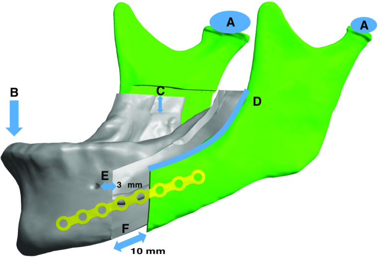
Background: The surgical treatment for mandibular repositioning using a bilateral sagittal split osteotomy (BSSO) favours the development of techniques that result in adequate repair and stability. In Puricelli's mandibular sagittal split osteotomy (PMSSO) proposal, the vertical lateral cut osteotomy is located in the interradicular space between the lower first molar and second premolar.
Objectives: This in silico study aimed to investigate the mechanical stability of PMSSO and compare it with the classical Obwegeser-Dal Pont technique for mandibular advancement.
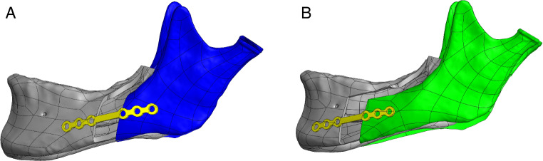
Materials and methods: A computational geometric model of the mandible was created in a virtual environment using computer-aided design (CAD) software. After reproducing the advancements, two test groups were developed: GTOD10, Obwegeser-Dal Pont osteotomy, and GTP10, Puricelli osteotomy, both simulating a 10-mm mandibular advancement, allowing for measuring the area of overlap between bone segments. With the geometric changes promoted by the osteotomy, boundary conditions of displacement and force were applied to a CAD software based on finite element analysis (FEA), allowing for quantitative and comparative analysis of the stress and vertical displacement of the mandible, mechanical measurements that may be associated with strength and stiffness.
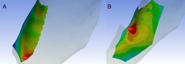
Results: A 17.48% higher stress was observed in the GTP10 group than in GTOD10. However, the region of highest stress in GTP10 was found in a part of the bone that was still intact and far from the area of fragility caused by lateral vertical osteotomy. In contrast, in GTOD10, the region with high stress was in a less resistant bone region. The GTP10 group showed a 28.73% lower displacement than GTOD10. The area of overlap between the proximal and distal segments of the mandible was 33.13% larger in the GTP10 than in the GTOD10 group.
Conclusion: The PMSSO method, performed in large mandibular advancements, keeps the point of highest stress away from the mandibular fragility zone. Considering the same amount of advancement, it also promotes less displacement and larger areas of bone overlap.
Clinical relevance: The results suggest that PMSSO, applied in large mandibular advancement, presents greater postoperative stability.
期刊介绍:
Head & Face Medicine is a multidisciplinary open access journal that publishes basic and clinical research concerning all aspects of cranial, facial and oral conditions.
The journal covers all aspects of cranial, facial and oral diseases and their management. It has been designed as a multidisciplinary journal for clinicians and researchers involved in the diagnostic and therapeutic aspects of diseases which affect the human head and face. The journal is wide-ranging, covering the development, aetiology, epidemiology and therapy of head and face diseases to the basic science that underlies these diseases. Management of head and face diseases includes all aspects of surgical and non-surgical treatments including psychopharmacological therapies.
 求助内容:
求助内容: 应助结果提醒方式:


