Praneeth Karempudi, Konrad Gras, Elias Amselem, Spartak Zikrin, Dvir Schirman, Johan Elf
{"title":"大肠杆菌细胞周期中染色体位点的三维定位和追踪。","authors":"Praneeth Karempudi, Konrad Gras, Elias Amselem, Spartak Zikrin, Dvir Schirman, Johan Elf","doi":"10.1038/s42003-024-07155-9","DOIUrl":null,"url":null,"abstract":"The intracellular position of genes may impact their expression, but it has not been possible to accurately measure the 3D position of chromosomal loci. In 2D, loci can be tracked using arrays of DNA-binding sites for transcription factors (TFs) fused with fluorescent proteins. However, the same 2D data can result from different 3D trajectories. Here, we have developed a deep learning method for super-resolved astigmatism-based 3D localization of chromosomal loci in live E. coli cells which enables a precision better than 61 nm at a signal-to-background ratio of ~4 on a heterogeneous cell background. Determining the spatial localization of chromosomal loci, we find that some loci are at the periphery of the nucleoid for large parts of the cell cycle. Analyses of individual trajectories reveal that these loci are subdiffusive both longitudinally (x) and radially (r), but that individual loci explore the full radial width on a minute time scale. The intracellular position of genes in the chromosome may impact their expression. To measure the 3D positions of chromosomal loci over the bacterial cell cycle, we have developed a combination of microfluidics, optics, and deep learning.","PeriodicalId":10552,"journal":{"name":"Communications Biology","volume":null,"pages":null},"PeriodicalIF":5.2000,"publicationDate":"2024-11-05","publicationTypes":"Journal Article","fieldsOfStudy":null,"isOpenAccess":false,"openAccessPdf":"https://www.ncbi.nlm.nih.gov/pmc/articles/PMC11538341/pdf/","citationCount":"0","resultStr":"{\"title\":\"Three-dimensional localization and tracking of chromosomal loci throughout the Escherichia coli cell cycle\",\"authors\":\"Praneeth Karempudi, Konrad Gras, Elias Amselem, Spartak Zikrin, Dvir Schirman, Johan Elf\",\"doi\":\"10.1038/s42003-024-07155-9\",\"DOIUrl\":null,\"url\":null,\"abstract\":\"The intracellular position of genes may impact their expression, but it has not been possible to accurately measure the 3D position of chromosomal loci. In 2D, loci can be tracked using arrays of DNA-binding sites for transcription factors (TFs) fused with fluorescent proteins. However, the same 2D data can result from different 3D trajectories. Here, we have developed a deep learning method for super-resolved astigmatism-based 3D localization of chromosomal loci in live E. coli cells which enables a precision better than 61 nm at a signal-to-background ratio of ~4 on a heterogeneous cell background. Determining the spatial localization of chromosomal loci, we find that some loci are at the periphery of the nucleoid for large parts of the cell cycle. Analyses of individual trajectories reveal that these loci are subdiffusive both longitudinally (x) and radially (r), but that individual loci explore the full radial width on a minute time scale. The intracellular position of genes in the chromosome may impact their expression. To measure the 3D positions of chromosomal loci over the bacterial cell cycle, we have developed a combination of microfluidics, optics, and deep learning.\",\"PeriodicalId\":10552,\"journal\":{\"name\":\"Communications Biology\",\"volume\":null,\"pages\":null},\"PeriodicalIF\":5.2000,\"publicationDate\":\"2024-11-05\",\"publicationTypes\":\"Journal Article\",\"fieldsOfStudy\":null,\"isOpenAccess\":false,\"openAccessPdf\":\"https://www.ncbi.nlm.nih.gov/pmc/articles/PMC11538341/pdf/\",\"citationCount\":\"0\",\"resultStr\":null,\"platform\":\"Semanticscholar\",\"paperid\":null,\"PeriodicalName\":\"Communications Biology\",\"FirstCategoryId\":\"99\",\"ListUrlMain\":\"https://www.nature.com/articles/s42003-024-07155-9\",\"RegionNum\":1,\"RegionCategory\":\"生物学\",\"ArticlePicture\":[],\"TitleCN\":null,\"AbstractTextCN\":null,\"PMCID\":null,\"EPubDate\":\"\",\"PubModel\":\"\",\"JCR\":\"Q1\",\"JCRName\":\"BIOLOGY\",\"Score\":null,\"Total\":0}","platform":"Semanticscholar","paperid":null,"PeriodicalName":"Communications Biology","FirstCategoryId":"99","ListUrlMain":"https://www.nature.com/articles/s42003-024-07155-9","RegionNum":1,"RegionCategory":"生物学","ArticlePicture":[],"TitleCN":null,"AbstractTextCN":null,"PMCID":null,"EPubDate":"","PubModel":"","JCR":"Q1","JCRName":"BIOLOGY","Score":null,"Total":0}
Three-dimensional localization and tracking of chromosomal loci throughout the Escherichia coli cell cycle
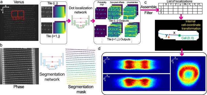
The intracellular position of genes may impact their expression, but it has not been possible to accurately measure the 3D position of chromosomal loci. In 2D, loci can be tracked using arrays of DNA-binding sites for transcription factors (TFs) fused with fluorescent proteins. However, the same 2D data can result from different 3D trajectories. Here, we have developed a deep learning method for super-resolved astigmatism-based 3D localization of chromosomal loci in live E. coli cells which enables a precision better than 61 nm at a signal-to-background ratio of ~4 on a heterogeneous cell background. Determining the spatial localization of chromosomal loci, we find that some loci are at the periphery of the nucleoid for large parts of the cell cycle. Analyses of individual trajectories reveal that these loci are subdiffusive both longitudinally (x) and radially (r), but that individual loci explore the full radial width on a minute time scale. The intracellular position of genes in the chromosome may impact their expression. To measure the 3D positions of chromosomal loci over the bacterial cell cycle, we have developed a combination of microfluidics, optics, and deep learning.
期刊介绍:
Communications Biology is an open access journal from Nature Research publishing high-quality research, reviews and commentary in all areas of the biological sciences. Research papers published by the journal represent significant advances bringing new biological insight to a specialized area of research.