Xiaohui Meng , Zhangchen Xia , Junwen Cheng , Yanbin Wang , Xueyong Ren , Liang He , Dan Liu
{"title":"评估麦角硫因与人血清白蛋白的结合机制:多光谱分析、分子对接和分子动力学模拟","authors":"Xiaohui Meng , Zhangchen Xia , Junwen Cheng , Yanbin Wang , Xueyong Ren , Liang He , Dan Liu","doi":"10.1016/j.saa.2024.125368","DOIUrl":null,"url":null,"abstract":"<div><div>Ergothioneine (EGT) has attracted great attention due to its extremely potent antioxidant properties, universally acknowledged as ‘longevity vitamin’. In order to comprehensive understanding of its pharmacodynamics and pharmacokinetics, the binding mechanism of EGT with human serum albumin (HSA) was clarified by cutting-edged multi-spectroscopic approaches and <em>in silico</em> molecular docking coupled with molecular dynamic simulation. Our fluorescence quenching results revealed that the binding of EGT to HSA was in a static quenching mode validated by the descending Stern–Volmer constant (<em>K</em><sub>sv</sub>) values (2.82, 2.36, 1.48 × 10<sup>4</sup> L mol<sup>−1</sup>) and biomolecular quenching rate constant (<em>K</em><sub>q</sub>) values (2.82, 2.36, 1.48 × 10<sup>12</sup> L mol<sup>−1</sup>) at 298 K, 305 K, and 310 K, respectively. van’t Hoff criterion revealed the combination of EGT with HSA was a spontaneous exothermic process (Δ<em>G</em> = −24.16 kJ mol<sup>−1</sup>) <em>via</em> hydrogen bonding and van der Waals force interactions (Δ<em>H</em> = −60.25 kJ mol<sup>−1</sup>, Δ<em>S</em> = −129.44 J mol<sup>−1</sup> K<sup>−1</sup>) at 310 K. The analysis of UV–vis absorption spectrum, synchronous fluorescence spectrum, three-dimensional fluorescence spectrum and circular dichroism indicated the addition of EGT affected the microenvironment of Trp214 and rearranged the structure of HSA. The binding replacement assay interpreted their binding site was near the subdomain IIA of HSA (Sudlow’s site I), which was intuitively exhibited by molecular docking. In addition of obvious van der Wall forces, attractive charge and Pi-alkyl interactions, the chiral betaine group (N<sup>+</sup>(CH<sub>3</sub><sup>+</sup>)<sub>3</sub>) in the side chain of EGT was inclined to form hydrogen bonds with Lys199, Ser287 and Arg257 in the hydrophobic cavity of albumin. Moreover, the dynamic simulation reinforced the equilibrium and stability of formed docking complex by four indicators (RMSD, RMSF, Rg, SASA) within 100 ns.</div></div>","PeriodicalId":433,"journal":{"name":"Spectrochimica Acta Part A: Molecular and Biomolecular Spectroscopy","volume":"327 ","pages":"Article 125368"},"PeriodicalIF":4.3000,"publicationDate":"2024-11-03","publicationTypes":"Journal Article","fieldsOfStudy":null,"isOpenAccess":false,"openAccessPdf":"","citationCount":"0","resultStr":"{\"title\":\"Assessment of the binding mechanism of ergothioneine to human serum albumin: Multi-spectroscopy, molecular docking and molecular dynamic simulation\",\"authors\":\"Xiaohui Meng , Zhangchen Xia , Junwen Cheng , Yanbin Wang , Xueyong Ren , Liang He , Dan Liu\",\"doi\":\"10.1016/j.saa.2024.125368\",\"DOIUrl\":null,\"url\":null,\"abstract\":\"<div><div>Ergothioneine (EGT) has attracted great attention due to its extremely potent antioxidant properties, universally acknowledged as ‘longevity vitamin’. In order to comprehensive understanding of its pharmacodynamics and pharmacokinetics, the binding mechanism of EGT with human serum albumin (HSA) was clarified by cutting-edged multi-spectroscopic approaches and <em>in silico</em> molecular docking coupled with molecular dynamic simulation. Our fluorescence quenching results revealed that the binding of EGT to HSA was in a static quenching mode validated by the descending Stern–Volmer constant (<em>K</em><sub>sv</sub>) values (2.82, 2.36, 1.48 × 10<sup>4</sup> L mol<sup>−1</sup>) and biomolecular quenching rate constant (<em>K</em><sub>q</sub>) values (2.82, 2.36, 1.48 × 10<sup>12</sup> L mol<sup>−1</sup>) at 298 K, 305 K, and 310 K, respectively. van’t Hoff criterion revealed the combination of EGT with HSA was a spontaneous exothermic process (Δ<em>G</em> = −24.16 kJ mol<sup>−1</sup>) <em>via</em> hydrogen bonding and van der Waals force interactions (Δ<em>H</em> = −60.25 kJ mol<sup>−1</sup>, Δ<em>S</em> = −129.44 J mol<sup>−1</sup> K<sup>−1</sup>) at 310 K. The analysis of UV–vis absorption spectrum, synchronous fluorescence spectrum, three-dimensional fluorescence spectrum and circular dichroism indicated the addition of EGT affected the microenvironment of Trp214 and rearranged the structure of HSA. The binding replacement assay interpreted their binding site was near the subdomain IIA of HSA (Sudlow’s site I), which was intuitively exhibited by molecular docking. In addition of obvious van der Wall forces, attractive charge and Pi-alkyl interactions, the chiral betaine group (N<sup>+</sup>(CH<sub>3</sub><sup>+</sup>)<sub>3</sub>) in the side chain of EGT was inclined to form hydrogen bonds with Lys199, Ser287 and Arg257 in the hydrophobic cavity of albumin. Moreover, the dynamic simulation reinforced the equilibrium and stability of formed docking complex by four indicators (RMSD, RMSF, Rg, SASA) within 100 ns.</div></div>\",\"PeriodicalId\":433,\"journal\":{\"name\":\"Spectrochimica Acta Part A: Molecular and Biomolecular Spectroscopy\",\"volume\":\"327 \",\"pages\":\"Article 125368\"},\"PeriodicalIF\":4.3000,\"publicationDate\":\"2024-11-03\",\"publicationTypes\":\"Journal Article\",\"fieldsOfStudy\":null,\"isOpenAccess\":false,\"openAccessPdf\":\"\",\"citationCount\":\"0\",\"resultStr\":null,\"platform\":\"Semanticscholar\",\"paperid\":null,\"PeriodicalName\":\"Spectrochimica Acta Part A: Molecular and Biomolecular Spectroscopy\",\"FirstCategoryId\":\"92\",\"ListUrlMain\":\"https://www.sciencedirect.com/science/article/pii/S1386142524015348\",\"RegionNum\":2,\"RegionCategory\":\"化学\",\"ArticlePicture\":[],\"TitleCN\":null,\"AbstractTextCN\":null,\"PMCID\":null,\"EPubDate\":\"\",\"PubModel\":\"\",\"JCR\":\"Q1\",\"JCRName\":\"SPECTROSCOPY\",\"Score\":null,\"Total\":0}","platform":"Semanticscholar","paperid":null,"PeriodicalName":"Spectrochimica Acta Part A: Molecular and Biomolecular Spectroscopy","FirstCategoryId":"92","ListUrlMain":"https://www.sciencedirect.com/science/article/pii/S1386142524015348","RegionNum":2,"RegionCategory":"化学","ArticlePicture":[],"TitleCN":null,"AbstractTextCN":null,"PMCID":null,"EPubDate":"","PubModel":"","JCR":"Q1","JCRName":"SPECTROSCOPY","Score":null,"Total":0}
Assessment of the binding mechanism of ergothioneine to human serum albumin: Multi-spectroscopy, molecular docking and molecular dynamic simulation
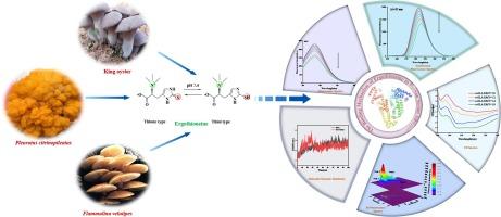
Ergothioneine (EGT) has attracted great attention due to its extremely potent antioxidant properties, universally acknowledged as ‘longevity vitamin’. In order to comprehensive understanding of its pharmacodynamics and pharmacokinetics, the binding mechanism of EGT with human serum albumin (HSA) was clarified by cutting-edged multi-spectroscopic approaches and in silico molecular docking coupled with molecular dynamic simulation. Our fluorescence quenching results revealed that the binding of EGT to HSA was in a static quenching mode validated by the descending Stern–Volmer constant (Ksv) values (2.82, 2.36, 1.48 × 10⁴ L mol−1) and biomolecular quenching rate constant (Kq) values (2.82, 2.36, 1.48 × 10¹² L mol−1) at 298 K, 305 K, and 310 K, respectively. van’t Hoff criterion revealed the combination of EGT with HSA was a spontaneous exothermic process (ΔG = −24.16 kJ mol−1) via hydrogen bonding and van der Waals force interactions (ΔH = −60.25 kJ mol−1, ΔS = −129.44 J mol−1 K−1) at 310 K. The analysis of UV–vis absorption spectrum, synchronous fluorescence spectrum, three-dimensional fluorescence spectrum and circular dichroism indicated the addition of EGT affected the microenvironment of Trp214 and rearranged the structure of HSA. The binding replacement assay interpreted their binding site was near the subdomain IIA of HSA (Sudlow’s site I), which was intuitively exhibited by molecular docking. In addition of obvious van der Wall forces, attractive charge and Pi-alkyl interactions, the chiral betaine group (N+(CH3+)3) in the side chain of EGT was inclined to form hydrogen bonds with Lys199, Ser287 and Arg257 in the hydrophobic cavity of albumin. Moreover, the dynamic simulation reinforced the equilibrium and stability of formed docking complex by four indicators (RMSD, RMSF, Rg, SASA) within 100 ns.
期刊介绍:
Spectrochimica Acta, Part A: Molecular and Biomolecular Spectroscopy (SAA) is an interdisciplinary journal which spans from basic to applied aspects of optical spectroscopy in chemistry, medicine, biology, and materials science.
The journal publishes original scientific papers that feature high-quality spectroscopic data and analysis. From the broad range of optical spectroscopies, the emphasis is on electronic, vibrational or rotational spectra of molecules, rather than on spectroscopy based on magnetic moments.
Criteria for publication in SAA are novelty, uniqueness, and outstanding quality. Routine applications of spectroscopic techniques and computational methods are not appropriate.
Topics of particular interest of Spectrochimica Acta Part A include, but are not limited to:
Spectroscopy and dynamics of bioanalytical, biomedical, environmental, and atmospheric sciences,
Novel experimental techniques or instrumentation for molecular spectroscopy,
Novel theoretical and computational methods,
Novel applications in photochemistry and photobiology,
Novel interpretational approaches as well as advances in data analysis based on electronic or vibrational spectroscopy.