{"title":"pDNA-Buforin II 对前列腺癌中 lncRNA PCA3、PCAT1、PRNCR1、GAS5 表达变化的影响","authors":"Fatemeh Dehkhodaei , Abbas Doosti","doi":"10.1016/j.adcanc.2024.100130","DOIUrl":null,"url":null,"abstract":"<div><h3>Background</h3><div>This work aims to analyze the alterations in the levels of <em>PCA3</em>, <em>PCAT1</em>, <em>PRNCR1</em>, and <em>GAS5</em> long non-coding RNAs (lncRNAs) after the activation of pDNA-buforin II in PC3 cancer cells.</div></div><div><h3>Materials and methods</h3><div>The synthetic nucleic acid sequence of buforin II was included in the pcDNA3.1(+) Mammalian Expression Plasmid. The accuracy of cloning was assessed by using PCR and enzyme digestion techniques. The vectors were transfected into cells utilizing LipofectamineTM2000. The PC3 cancer cells were evaluated using flow cytometry and wound healing analysis. The expression levels of lncRNAs and apoptotic genes were assessed utilizing real-time PCR, with a significance threshold of P < 0.05.</div></div><div><h3>Results</h3><div>The recombinant plasmid containing the pDNA-buforin II vector was successfully generated, and the gene sequence demonstrated complete uniformity (100 % similarity) with the buforin II gene. The transfection efficiency of PC3 cells was 79 %. The results are quantified utilizing the growth inhibition 50 % (GI50) parameter, representing the concentration of pDNA-buforin II required to halt 50 % of cell growth. The percentages of early apoptosis, late apoptosis, necrosis, and viable PC3 cells in the pDNA-buforin II group were 23.30 %, 12.70 %, 3.9 %, and 60.10 %, respectively. The RT-PCR study demonstrated that the presence of pDNA-buforin II in PC3 cells decreased the transcription of <em>PCA3</em>, <em>PCAT1</em>, and <em>PRNCR1</em> lncRNAs compared to the control group treated with PBS. Furthermore, it enhanced the transcription of <em>GAS5</em> lncRNA. The findings demonstrated a significant upregulation of transcription factors in programmed cell death after treatment with pDNA-buforin II (∗∗P < 0.01).</div></div><div><h3>Conclusions</h3><div>According to the results of this study, it can be inferred that pDNA-buforin II can modify the transcription of genes in PC3 cancer cells, specifically about lncRNAs involved in cell apoptotic pathways. The pDNA-buforin II molecule has promising anticancer capabilities and can trigger apoptosis in cells.</div></div>","PeriodicalId":72083,"journal":{"name":"Advances in cancer biology - metastasis","volume":null,"pages":null},"PeriodicalIF":2.0000,"publicationDate":"2024-10-30","publicationTypes":"Journal Article","fieldsOfStudy":null,"isOpenAccess":false,"openAccessPdf":"","citationCount":"0","resultStr":"{\"title\":\"The effect of pDNA-Buforin II on the expression changes of lncRNAs PCA3, PCAT1, PRNCR1, GAS5 in prostate cancer\",\"authors\":\"Fatemeh Dehkhodaei , Abbas Doosti\",\"doi\":\"10.1016/j.adcanc.2024.100130\",\"DOIUrl\":null,\"url\":null,\"abstract\":\"<div><h3>Background</h3><div>This work aims to analyze the alterations in the levels of <em>PCA3</em>, <em>PCAT1</em>, <em>PRNCR1</em>, and <em>GAS5</em> long non-coding RNAs (lncRNAs) after the activation of pDNA-buforin II in PC3 cancer cells.</div></div><div><h3>Materials and methods</h3><div>The synthetic nucleic acid sequence of buforin II was included in the pcDNA3.1(+) Mammalian Expression Plasmid. The accuracy of cloning was assessed by using PCR and enzyme digestion techniques. The vectors were transfected into cells utilizing LipofectamineTM2000. The PC3 cancer cells were evaluated using flow cytometry and wound healing analysis. The expression levels of lncRNAs and apoptotic genes were assessed utilizing real-time PCR, with a significance threshold of P < 0.05.</div></div><div><h3>Results</h3><div>The recombinant plasmid containing the pDNA-buforin II vector was successfully generated, and the gene sequence demonstrated complete uniformity (100 % similarity) with the buforin II gene. The transfection efficiency of PC3 cells was 79 %. The results are quantified utilizing the growth inhibition 50 % (GI50) parameter, representing the concentration of pDNA-buforin II required to halt 50 % of cell growth. The percentages of early apoptosis, late apoptosis, necrosis, and viable PC3 cells in the pDNA-buforin II group were 23.30 %, 12.70 %, 3.9 %, and 60.10 %, respectively. The RT-PCR study demonstrated that the presence of pDNA-buforin II in PC3 cells decreased the transcription of <em>PCA3</em>, <em>PCAT1</em>, and <em>PRNCR1</em> lncRNAs compared to the control group treated with PBS. Furthermore, it enhanced the transcription of <em>GAS5</em> lncRNA. The findings demonstrated a significant upregulation of transcription factors in programmed cell death after treatment with pDNA-buforin II (∗∗P < 0.01).</div></div><div><h3>Conclusions</h3><div>According to the results of this study, it can be inferred that pDNA-buforin II can modify the transcription of genes in PC3 cancer cells, specifically about lncRNAs involved in cell apoptotic pathways. The pDNA-buforin II molecule has promising anticancer capabilities and can trigger apoptosis in cells.</div></div>\",\"PeriodicalId\":72083,\"journal\":{\"name\":\"Advances in cancer biology - metastasis\",\"volume\":null,\"pages\":null},\"PeriodicalIF\":2.0000,\"publicationDate\":\"2024-10-30\",\"publicationTypes\":\"Journal Article\",\"fieldsOfStudy\":null,\"isOpenAccess\":false,\"openAccessPdf\":\"\",\"citationCount\":\"0\",\"resultStr\":null,\"platform\":\"Semanticscholar\",\"paperid\":null,\"PeriodicalName\":\"Advances in cancer biology - metastasis\",\"FirstCategoryId\":\"1085\",\"ListUrlMain\":\"https://www.sciencedirect.com/science/article/pii/S2667394024000170\",\"RegionNum\":0,\"RegionCategory\":null,\"ArticlePicture\":[],\"TitleCN\":null,\"AbstractTextCN\":null,\"PMCID\":null,\"EPubDate\":\"\",\"PubModel\":\"\",\"JCR\":\"Q3\",\"JCRName\":\"ONCOLOGY\",\"Score\":null,\"Total\":0}","platform":"Semanticscholar","paperid":null,"PeriodicalName":"Advances in cancer biology - metastasis","FirstCategoryId":"1085","ListUrlMain":"https://www.sciencedirect.com/science/article/pii/S2667394024000170","RegionNum":0,"RegionCategory":null,"ArticlePicture":[],"TitleCN":null,"AbstractTextCN":null,"PMCID":null,"EPubDate":"","PubModel":"","JCR":"Q3","JCRName":"ONCOLOGY","Score":null,"Total":0}
引用次数: 0
摘要
背景本研究旨在分析 pDNA-buforin II 激活 PC3 癌细胞后 PCA3、PCAT1、PRNCR1 和 GAS5 长非编码 RNA(lncRNA)水平的变化。利用 PCR 和酶切技术评估了克隆的准确性。利用 LipofectamineTM2000 将载体转染到细胞中。使用流式细胞仪和伤口愈合分析对 PC3 癌细胞进行评估。结果pDNA-buforin II载体重组质粒成功产生,其基因序列与buforin II基因完全一致(100%相似)。PC3 细胞的转染效率为 79%。转染结果用生长抑制 50%(GI50)参数进行量化,该参数代表阻止 50% 细胞生长所需的 pDNA-buforin II 浓度。pDNA-buforin II 组早期凋亡、晚期凋亡、坏死和存活 PC3 细胞的百分比分别为 23.30%、12.70%、3.9% 和 60.10%。RT-PCR 研究表明,与用 PBS 处理的对照组相比,pDNA-buforin II 在 PC3 细胞中的存在降低了 PCA3、PCAT1 和 PRNCR1 lncRNA 的转录。此外,它还增强了 GAS5 lncRNA 的转录。结论根据本研究的结果,可以推断 pDNA-buforin II 可以改变 PC3 癌细胞中基因的转录,特别是参与细胞凋亡通路的 lncRNA。pDNA-buforin II 分子具有很好的抗癌能力,能引发细胞凋亡。
The effect of pDNA-Buforin II on the expression changes of lncRNAs PCA3, PCAT1, PRNCR1, GAS5 in prostate cancer
Background
This work aims to analyze the alterations in the levels of PCA3, PCAT1, PRNCR1, and GAS5 long non-coding RNAs (lncRNAs) after the activation of pDNA-buforin II in PC3 cancer cells.
Materials and methods
The synthetic nucleic acid sequence of buforin II was included in the pcDNA3.1(+) Mammalian Expression Plasmid. The accuracy of cloning was assessed by using PCR and enzyme digestion techniques. The vectors were transfected into cells utilizing LipofectamineTM2000. The PC3 cancer cells were evaluated using flow cytometry and wound healing analysis. The expression levels of lncRNAs and apoptotic genes were assessed utilizing real-time PCR, with a significance threshold of P < 0.05.
Results
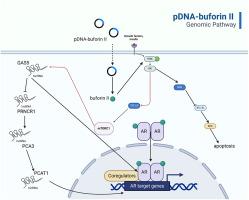
The recombinant plasmid containing the pDNA-buforin II vector was successfully generated, and the gene sequence demonstrated complete uniformity (100 % similarity) with the buforin II gene. The transfection efficiency of PC3 cells was 79 %. The results are quantified utilizing the growth inhibition 50 % (GI50) parameter, representing the concentration of pDNA-buforin II required to halt 50 % of cell growth. The percentages of early apoptosis, late apoptosis, necrosis, and viable PC3 cells in the pDNA-buforin II group were 23.30 %, 12.70 %, 3.9 %, and 60.10 %, respectively. The RT-PCR study demonstrated that the presence of pDNA-buforin II in PC3 cells decreased the transcription of PCA3, PCAT1, and PRNCR1 lncRNAs compared to the control group treated with PBS. Furthermore, it enhanced the transcription of GAS5 lncRNA. The findings demonstrated a significant upregulation of transcription factors in programmed cell death after treatment with pDNA-buforin II (∗∗P < 0.01).
Conclusions
According to the results of this study, it can be inferred that pDNA-buforin II can modify the transcription of genes in PC3 cancer cells, specifically about lncRNAs involved in cell apoptotic pathways. The pDNA-buforin II molecule has promising anticancer capabilities and can trigger apoptosis in cells.