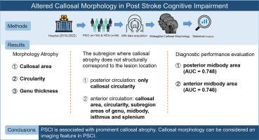
中风后认知障碍中的胼胝体形态改变。

IF 2.7 4区 医学 Q3 NEUROSCIENCES
Xiaoli Zhou , Huan Li , Tao Li, Zhao Ruan, Xiaohui Chen, Xitong Liu, Lei Gao, Haibo Xu
{"title":"中风后认知障碍中的胼胝体形态改变。","authors":"Xiaoli Zhou ,&nbsp;Huan Li ,&nbsp;Tao Li,&nbsp;Zhao Ruan,&nbsp;Xiaohui Chen,&nbsp;Xitong Liu,&nbsp;Lei Gao,&nbsp;Haibo Xu","doi":"10.1016/j.brainres.2024.149307","DOIUrl":null,"url":null,"abstract":"<div><div>Stroke is the second leading cause of death and cognitive impairment. Post-stroke cognitive impairment (PSCI) is one of the most common sequelae among stroke survivors, yet its underlying neural mechanisms remain largely unclear. The corpus callosum (CC) plays a crucial role in interhemispheric integration and hemispheric segregation, with changes in CC morphology potentially overlapping with the spectrum of PSCI. This study aimed to investigate the morphological changes in the CC and their diagnostic value in PSCI patients. Structural MRI, neurobehavioral, and clinical data were collected from 104 PSCI patients and 54 demographically matched healthy controls. Significant reductions in CC area, circularity, and genu thickness were observed in PSCI patients, with these changes strongly correlating with global cognitive function. Subgroup analysis revealed that CC circularity significantly decreased when lesions were located in the posterior circulation, while both CC area and circularity markedly decreased with anterior circulation lesions. Receiver Operating Characteristic analyses identified the midbody areas of the CC as having high diagnostic value, with area under the curve values of 0.748 and 0.746, respectively. Further validation analyses suggest that the transcallosal fibers in these CC subregions are connected to the premotor, dorsal attention, and frontoparietal system. These findings show that areal CC atrophy in PSCI patients, particularly in regions with transcallosal connections to the premotor cortex and frontoparietal network, parallels global cognitive impairment. This suggests that CC morphology may serve as a potential imaging marker for the diagnosis and prognosis of PSCI.</div></div>","PeriodicalId":9083,"journal":{"name":"Brain Research","volume":"1847 ","pages":"Article 149307"},"PeriodicalIF":2.7000,"publicationDate":"2024-11-02","publicationTypes":"Journal Article","fieldsOfStudy":null,"isOpenAccess":false,"openAccessPdf":"","citationCount":"0","resultStr":"{\"title\":\"Altered callosal morphology in post-stroke cognitive impairment\",\"authors\":\"Xiaoli Zhou ,&nbsp;Huan Li ,&nbsp;Tao Li,&nbsp;Zhao Ruan,&nbsp;Xiaohui Chen,&nbsp;Xitong Liu,&nbsp;Lei Gao,&nbsp;Haibo Xu\",\"doi\":\"10.1016/j.brainres.2024.149307\",\"DOIUrl\":null,\"url\":null,\"abstract\":\"<div><div>Stroke is the second leading cause of death and cognitive impairment. Post-stroke cognitive impairment (PSCI) is one of the most common sequelae among stroke survivors, yet its underlying neural mechanisms remain largely unclear. The corpus callosum (CC) plays a crucial role in interhemispheric integration and hemispheric segregation, with changes in CC morphology potentially overlapping with the spectrum of PSCI. This study aimed to investigate the morphological changes in the CC and their diagnostic value in PSCI patients. Structural MRI, neurobehavioral, and clinical data were collected from 104 PSCI patients and 54 demographically matched healthy controls. Significant reductions in CC area, circularity, and genu thickness were observed in PSCI patients, with these changes strongly correlating with global cognitive function. Subgroup analysis revealed that CC circularity significantly decreased when lesions were located in the posterior circulation, while both CC area and circularity markedly decreased with anterior circulation lesions. Receiver Operating Characteristic analyses identified the midbody areas of the CC as having high diagnostic value, with area under the curve values of 0.748 and 0.746, respectively. Further validation analyses suggest that the transcallosal fibers in these CC subregions are connected to the premotor, dorsal attention, and frontoparietal system. These findings show that areal CC atrophy in PSCI patients, particularly in regions with transcallosal connections to the premotor cortex and frontoparietal network, parallels global cognitive impairment. This suggests that CC morphology may serve as a potential imaging marker for the diagnosis and prognosis of PSCI.</div></div>\",\"PeriodicalId\":9083,\"journal\":{\"name\":\"Brain Research\",\"volume\":\"1847 \",\"pages\":\"Article 149307\"},\"PeriodicalIF\":2.7000,\"publicationDate\":\"2024-11-02\",\"publicationTypes\":\"Journal Article\",\"fieldsOfStudy\":null,\"isOpenAccess\":false,\"openAccessPdf\":\"\",\"citationCount\":\"0\",\"resultStr\":null,\"platform\":\"Semanticscholar\",\"paperid\":null,\"PeriodicalName\":\"Brain Research\",\"FirstCategoryId\":\"3\",\"ListUrlMain\":\"https://www.sciencedirect.com/science/article/pii/S0006899324005614\",\"RegionNum\":4,\"RegionCategory\":\"医学\",\"ArticlePicture\":[],\"TitleCN\":null,\"AbstractTextCN\":null,\"PMCID\":null,\"EPubDate\":\"\",\"PubModel\":\"\",\"JCR\":\"Q3\",\"JCRName\":\"NEUROSCIENCES\",\"Score\":null,\"Total\":0}","platform":"Semanticscholar","paperid":null,"PeriodicalName":"Brain Research","FirstCategoryId":"3","ListUrlMain":"https://www.sciencedirect.com/science/article/pii/S0006899324005614","RegionNum":4,"RegionCategory":"医学","ArticlePicture":[],"TitleCN":null,"AbstractTextCN":null,"PMCID":null,"EPubDate":"","PubModel":"","JCR":"Q3","JCRName":"NEUROSCIENCES","Score":null,"Total":0}
引用次数: 0

摘要

中风是导致死亡和认知障碍的第二大原因。脑卒中后认知障碍(PSCI)是脑卒中幸存者最常见的后遗症之一,但其潜在的神经机制在很大程度上仍不清楚。胼胝体(CC)在半球间整合和半球分离中起着至关重要的作用,CC形态学的变化可能与PSCI的范围重叠。本研究旨在探讨CC的形态学变化及其在PSCI患者中的诊断价值。研究收集了104名PSCI患者和54名人口统计学匹配的健康对照者的结构磁共振成像、神经行为和临床数据。在PSCI患者中观察到CC面积、圆度和真皮厚度显著减少,这些变化与整体认知功能密切相关。亚组分析显示,当病变位于后循环时,CC的圆度明显下降,而当病变位于前循环时,CC面积和圆度均明显下降。接收者操作特征分析表明,CC的中体区域具有很高的诊断价值,其曲线下面积值分别为0.748和0.746。进一步的验证分析表明,这些CC亚区的跨胼胝体纤维与前运动、背侧注意和顶叶前部系统相连。这些研究结果表明,PSCI 患者的 CC 肢体萎缩,尤其是在与前运动皮层和额顶叶网络有跨胼胝体连接的区域,与整体认知障碍并行。这表明,CC形态学可作为诊断和预后PSCI的潜在影像标记。
本文章由计算机程序翻译,如有差异,请以英文原文为准。

Altered callosal morphology in post-stroke cognitive impairment

Altered callosal morphology in post-stroke cognitive impairment
Stroke is the second leading cause of death and cognitive impairment. Post-stroke cognitive impairment (PSCI) is one of the most common sequelae among stroke survivors, yet its underlying neural mechanisms remain largely unclear. The corpus callosum (CC) plays a crucial role in interhemispheric integration and hemispheric segregation, with changes in CC morphology potentially overlapping with the spectrum of PSCI. This study aimed to investigate the morphological changes in the CC and their diagnostic value in PSCI patients. Structural MRI, neurobehavioral, and clinical data were collected from 104 PSCI patients and 54 demographically matched healthy controls. Significant reductions in CC area, circularity, and genu thickness were observed in PSCI patients, with these changes strongly correlating with global cognitive function. Subgroup analysis revealed that CC circularity significantly decreased when lesions were located in the posterior circulation, while both CC area and circularity markedly decreased with anterior circulation lesions. Receiver Operating Characteristic analyses identified the midbody areas of the CC as having high diagnostic value, with area under the curve values of 0.748 and 0.746, respectively. Further validation analyses suggest that the transcallosal fibers in these CC subregions are connected to the premotor, dorsal attention, and frontoparietal system. These findings show that areal CC atrophy in PSCI patients, particularly in regions with transcallosal connections to the premotor cortex and frontoparietal network, parallels global cognitive impairment. This suggests that CC morphology may serve as a potential imaging marker for the diagnosis and prognosis of PSCI.
求助全文
通过发布文献求助,成功后即可免费获取论文全文。 去求助
来源期刊
Brain Research
Brain Research 医学-神经科学
CiteScore
5.90
自引率
3.40%
发文量
268
审稿时长
47 days
期刊介绍: An international multidisciplinary journal devoted to fundamental research in the brain sciences. Brain Research publishes papers reporting interdisciplinary investigations of nervous system structure and function that are of general interest to the international community of neuroscientists. As is evident from the journals name, its scope is broad, ranging from cellular and molecular studies through systems neuroscience, cognition and disease. Invited reviews are also published; suggestions for and inquiries about potential reviews are welcomed. With the appearance of the final issue of the 2011 subscription, Vol. 67/1-2 (24 June 2011), Brain Research Reviews has ceased publication as a distinct journal separate from Brain Research. Review articles accepted for Brain Research are now published in that journal.
×
引用
GB/T 7714-2015
复制
MLA
复制
APA
复制
导出至
BibTeX EndNote RefMan NoteFirst NoteExpress
×
提示
您的信息不完整,为了账户安全,请先补充。
现在去补充
×
提示
您因"违规操作"
具体请查看互助需知
我知道了
×
提示
确定
请完成安全验证×
copy
已复制链接
快去分享给好友吧!
我知道了
右上角分享
点击右上角分享
0
联系我们:info@booksci.cn Book学术提供免费学术资源搜索服务,方便国内外学者检索中英文文献。致力于提供最便捷和优质的服务体验。 Copyright © 2023 布克学术 All rights reserved.
京ICP备2023020795号-1
ghs 京公网安备 11010802042870号
Book学术文献互助
Book学术文献互助群
群 号:481959085
Book学术官方微信