用于心脏瓣膜修复的 3D 打印胶原支架
Q3 Medicine
引用次数: 0
摘要
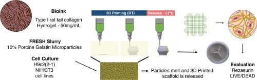
三维(3D)生物打印已成为开发包括心脏瓣膜在内的功能性组织和器官的一种前景广阔的方法。在本研究中,我们研究了三维打印胶原支架与 H9c2(2-1) 和 NIH/3T3 细胞的相互作用,以改进心脏瓣膜修复策略。我们从大鼠尾部提取了 I 型胶原蛋白,使用 SDS-PAGE 和拉曼光谱对其进行了表征,并将其用作三维打印的生物材料墨水。对其流变特性进行了评估。FRESH 技术用于支持打印的构建体。还进行了体外评估,以确定支架内的细胞活力和分布情况。这些结果表明,I 型胶原蛋白的提取和表征取得了成功,其流变特性适合用于三维生物打印。打印出的胶原支架支持 H9c2(2-1)和 NIH/3T3 细胞的生长和分布,这表明它们在心脏瓣膜修复中具有潜在的应用价值。这项研究强调了胶原蛋白作为一种生物材料在三维生物打印中的重要性,并为开发先进的心脏瓣膜修复策略提供了启示。本文章由计算机程序翻译,如有差异,请以英文原文为准。
3D printed collagen scaffold for heart valve repair
Three-dimensional (3D) bioprinting has emerged as a promising approach for the development of functional tissues and organs, including the heart valves. In this study, we investigated the interaction of 3D printed collagen scaffolds with H9c2(2–1) and NIH/3T3 cells to improve heart-valve repair strategies. Type I collagen was extracted from rat tails, characterized using SDS-PAGE and Raman spectroscopy, and used as a biomaterial ink for 3D printing. The rheological properties were evaluated. The FRESH technique was used to support the printed construct. In vitro assessments were performed to determine the cell viability and distribution within the scaffold. These results demonstrated the successful extraction and characterization of Type I collagen, which exhibited suitable rheological properties for 3D bioprinting. The printed collagen scaffolds supported the growth and distribution of H9c2(2–1) and NIH/3T3 cells, indicating their potential application in heart valve repair. This study highlights the importance of collagen as a biomaterial in 3D bioprinting and provides insights into the development of advanced strategies for heart valve repair.
求助全文
通过发布文献求助,成功后即可免费获取论文全文。
去求助
来源期刊

Annals of 3D printed medicine
Medicine and Dentistry (General), Materials Science (General)
CiteScore
4.70
自引率
0.00%
发文量
0
审稿时长
131 days
 求助内容:
求助内容: 应助结果提醒方式:


