Guilherme Gonçalves da Cruz, Roberta de Oliveira Alves, Caroline Garcia Orsi, André Luis Faria-E-Silva, Suzane Cristina Pigossi, Priscilla Barbosa Ferreira Soares
{"title":"牙周器械对牙齿结构暴露于电离辐射的影响:体外研究。","authors":"Guilherme Gonçalves da Cruz, Roberta de Oliveira Alves, Caroline Garcia Orsi, André Luis Faria-E-Silva, Suzane Cristina Pigossi, Priscilla Barbosa Ferreira Soares","doi":"10.1590/0103-6440202405763","DOIUrl":null,"url":null,"abstract":"<p><p>This study aimed to evaluate the effects of different scaling protocols on the morphology and roughness of the root dentin substrate exposure to ionizing radiation. One hundred and thirty extracted bovine incisors were randomly divided into two groups (n=65): non-irradiated (NIR) and irradiated (IR). Each group was initially subdivided into three subgroups according to the type of non-surgical periodontal protocol: NIT: no instrumentation; HS: hand scaling with 15 apical-coronal instrument movements; US: ultrasonic scaling with 15 apical-coronal cycles. Subsequently, all samples were subjected to the prophylaxis protocol, being subdivided into the following groups: NIT/PP: prophylaxis with a fine prophylactic paste using a rubber cup for 15 seconds; HS/PP: Hand scaling followed by the prophylaxis protocol; US/PP: Ultrasonic scaling followed by the prophylaxis protocol. The roughness of the root dentin surface was measured with a profilometer (Ra/Rz - μm), and the morphology of the dentin surfaces was analyzed using scanning electron microscopy (SEM). The analyses were conducted before and after the prophylaxis protocol. In the absence of prophylaxis, the roughest surfaces were observed after ultrasonic instrumentation followed by hand instrumentation for both IR and NIR groups. No difference in Ra and RZ values between HS/PP and US/PP was observed for both substrates. For the IR group, the prophylaxis resulted in similar Ra and RZ values for both instrumentation groups in comparison to no instrumentation. Ordinal logistic regression showed that both HS and US resulted in higher scores than NIT, irrespective of IR presence. In conclusion, the IR showed a rougher root surface for both HS and US in comparison to NIR. However, the prophylaxis procedure significantly reduced the roughness of root surfaces after both instrumentation procedures.</p>","PeriodicalId":101363,"journal":{"name":"Brazilian dental journal","volume":"35 ","pages":"e245763"},"PeriodicalIF":0.0000,"publicationDate":"2024-10-28","publicationTypes":"Journal Article","fieldsOfStudy":null,"isOpenAccess":false,"openAccessPdf":"https://www.ncbi.nlm.nih.gov/pmc/articles/PMC11520504/pdf/","citationCount":"0","resultStr":"{\"title\":\"Effect of Periodontal Instrumentation on Tooth Structure Exposure to Ionizing Radiation: In Vitro Study.\",\"authors\":\"Guilherme Gonçalves da Cruz, Roberta de Oliveira Alves, Caroline Garcia Orsi, André Luis Faria-E-Silva, Suzane Cristina Pigossi, Priscilla Barbosa Ferreira Soares\",\"doi\":\"10.1590/0103-6440202405763\",\"DOIUrl\":null,\"url\":null,\"abstract\":\"<p><p>This study aimed to evaluate the effects of different scaling protocols on the morphology and roughness of the root dentin substrate exposure to ionizing radiation. One hundred and thirty extracted bovine incisors were randomly divided into two groups (n=65): non-irradiated (NIR) and irradiated (IR). Each group was initially subdivided into three subgroups according to the type of non-surgical periodontal protocol: NIT: no instrumentation; HS: hand scaling with 15 apical-coronal instrument movements; US: ultrasonic scaling with 15 apical-coronal cycles. Subsequently, all samples were subjected to the prophylaxis protocol, being subdivided into the following groups: NIT/PP: prophylaxis with a fine prophylactic paste using a rubber cup for 15 seconds; HS/PP: Hand scaling followed by the prophylaxis protocol; US/PP: Ultrasonic scaling followed by the prophylaxis protocol. The roughness of the root dentin surface was measured with a profilometer (Ra/Rz - μm), and the morphology of the dentin surfaces was analyzed using scanning electron microscopy (SEM). The analyses were conducted before and after the prophylaxis protocol. In the absence of prophylaxis, the roughest surfaces were observed after ultrasonic instrumentation followed by hand instrumentation for both IR and NIR groups. No difference in Ra and RZ values between HS/PP and US/PP was observed for both substrates. For the IR group, the prophylaxis resulted in similar Ra and RZ values for both instrumentation groups in comparison to no instrumentation. Ordinal logistic regression showed that both HS and US resulted in higher scores than NIT, irrespective of IR presence. In conclusion, the IR showed a rougher root surface for both HS and US in comparison to NIR. However, the prophylaxis procedure significantly reduced the roughness of root surfaces after both instrumentation procedures.</p>\",\"PeriodicalId\":101363,\"journal\":{\"name\":\"Brazilian dental journal\",\"volume\":\"35 \",\"pages\":\"e245763\"},\"PeriodicalIF\":0.0000,\"publicationDate\":\"2024-10-28\",\"publicationTypes\":\"Journal Article\",\"fieldsOfStudy\":null,\"isOpenAccess\":false,\"openAccessPdf\":\"https://www.ncbi.nlm.nih.gov/pmc/articles/PMC11520504/pdf/\",\"citationCount\":\"0\",\"resultStr\":null,\"platform\":\"Semanticscholar\",\"paperid\":null,\"PeriodicalName\":\"Brazilian dental journal\",\"FirstCategoryId\":\"1085\",\"ListUrlMain\":\"https://doi.org/10.1590/0103-6440202405763\",\"RegionNum\":0,\"RegionCategory\":null,\"ArticlePicture\":[],\"TitleCN\":null,\"AbstractTextCN\":null,\"PMCID\":null,\"EPubDate\":\"2024/1/1 0:00:00\",\"PubModel\":\"eCollection\",\"JCR\":\"\",\"JCRName\":\"\",\"Score\":null,\"Total\":0}","platform":"Semanticscholar","paperid":null,"PeriodicalName":"Brazilian dental journal","FirstCategoryId":"1085","ListUrlMain":"https://doi.org/10.1590/0103-6440202405763","RegionNum":0,"RegionCategory":null,"ArticlePicture":[],"TitleCN":null,"AbstractTextCN":null,"PMCID":null,"EPubDate":"2024/1/1 0:00:00","PubModel":"eCollection","JCR":"","JCRName":"","Score":null,"Total":0}
引用次数: 0
摘要
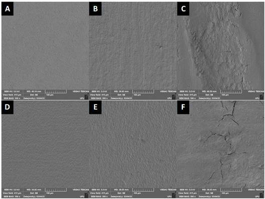
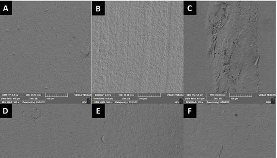
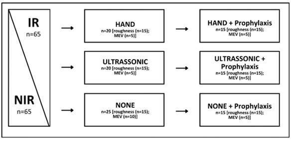
本研究旨在评估不同洗牙方案对暴露于电离辐射的牙根牙本质基底的形态和粗糙度的影响。将 130 颗拔出的牛门牙随机分为两组(n=65):非辐照组(NIR)和辐照组(IR)。根据非手术牙周治疗方案的类型,每组又分为三个亚组:NIT:无器械操作;HS:手洗牙,15 次根尖-冠状沟器械运动;US:超声波洗牙,15 次根尖-冠状沟循环。随后,所有样本都接受了预防方案,并被细分为以下几组:NIT/PP:使用橡皮杯在15秒钟内用精细的预防性糊剂进行预防;HS/PP:手工洗牙,然后进行预防性治疗;US/PP:超声波洗牙,然后进行预防性治疗。使用轮廓仪测量牙根牙本质表面的粗糙度(Ra/Rz - μm),并使用扫描电子显微镜(SEM)分析牙本质表面的形态。这些分析分别在预防方案前后进行。在没有预防措施的情况下,红外线组和近红外组的牙本质表面在超声波器械操作后最粗糙,然后是手工器械操作。对于两种基底,HS/PP 和 US/PP 的 Ra 值和 RZ 值均无差异。对于 IR 组,预防性治疗导致两组器械处理后的 Ra 值和 RZ 值与未进行器械处理时相似。顺序逻辑回归显示,无论是否存在 IR,HS 和 US 的评分均高于 NIT。总之,与 NIR 相比,HS 和 US 的 IR 显示牙根表面更粗糙。然而,预防程序显著降低了两种器械操作后牙根表面的粗糙度。
Effect of Periodontal Instrumentation on Tooth Structure Exposure to Ionizing Radiation: In Vitro Study.
This study aimed to evaluate the effects of different scaling protocols on the morphology and roughness of the root dentin substrate exposure to ionizing radiation. One hundred and thirty extracted bovine incisors were randomly divided into two groups (n=65): non-irradiated (NIR) and irradiated (IR). Each group was initially subdivided into three subgroups according to the type of non-surgical periodontal protocol: NIT: no instrumentation; HS: hand scaling with 15 apical-coronal instrument movements; US: ultrasonic scaling with 15 apical-coronal cycles. Subsequently, all samples were subjected to the prophylaxis protocol, being subdivided into the following groups: NIT/PP: prophylaxis with a fine prophylactic paste using a rubber cup for 15 seconds; HS/PP: Hand scaling followed by the prophylaxis protocol; US/PP: Ultrasonic scaling followed by the prophylaxis protocol. The roughness of the root dentin surface was measured with a profilometer (Ra/Rz - μm), and the morphology of the dentin surfaces was analyzed using scanning electron microscopy (SEM). The analyses were conducted before and after the prophylaxis protocol. In the absence of prophylaxis, the roughest surfaces were observed after ultrasonic instrumentation followed by hand instrumentation for both IR and NIR groups. No difference in Ra and RZ values between HS/PP and US/PP was observed for both substrates. For the IR group, the prophylaxis resulted in similar Ra and RZ values for both instrumentation groups in comparison to no instrumentation. Ordinal logistic regression showed that both HS and US resulted in higher scores than NIT, irrespective of IR presence. In conclusion, the IR showed a rougher root surface for both HS and US in comparison to NIR. However, the prophylaxis procedure significantly reduced the roughness of root surfaces after both instrumentation procedures.

 求助内容:
求助内容: 应助结果提醒方式:


