SHAPE:用于交互式三维照片标记和患者报告的视觉计算管道,以评估颅骨发育不良症

IF 2.5 4区 计算机科学 Q2 COMPUTER SCIENCE, SOFTWARE ENGINEERING
Carsten Görg , Connor Elkhill , Jasmine Chaij , Kristin Royalty , Phuong D. Nguyen , Brooke French , Ines A. Cruz-Guerrero , Antonio R. Porras
{"title":"SHAPE:用于交互式三维照片标记和患者报告的视觉计算管道,以评估颅骨发育不良症","authors":"Carsten Görg ,&nbsp;Connor Elkhill ,&nbsp;Jasmine Chaij ,&nbsp;Kristin Royalty ,&nbsp;Phuong D. Nguyen ,&nbsp;Brooke French ,&nbsp;Ines A. Cruz-Guerrero ,&nbsp;Antonio R. Porras","doi":"10.1016/j.cag.2024.104056","DOIUrl":null,"url":null,"abstract":"<div><div>3D photogrammetry is a cost-effective, non-invasive imaging modality that does not require the use of ionizing radiation or sedation. Therefore, it is specifically valuable in pediatrics and is used to support the diagnosis and longitudinal study of craniofacial developmental pathologies such as craniosynostosis — the premature fusion of one or more cranial sutures resulting in local cranial growth restrictions and cranial malformations. Analysis of 3D photogrammetry requires the identification of craniofacial landmarks to segment the head surface and compute metrics to quantify anomalies. Unfortunately, commercial 3D photogrammetry software requires intensive manual landmark placements, which is time-consuming and prone to errors. We designed and implemented SHAPE, a System for Head-shape Analysis and Pediatric Evaluation. It integrates our previously developed automated landmarking method in a visual computing pipeline to evaluate a patient’s 3D photogram while allowing for manual confirmation and correction. It also automatically computes advanced metrics to quantify craniofacial anomalies and automatically creates a report that can be uploaded to the patient’s electronic health record. We conducted a user study with a professional clinical photographer to compare SHAPE to the existing clinical workflow. We found that SHAPE allows for the evaluation of a craniofacial 3D photogram more than three times faster than the current clinical workflow (<span><math><mrow><mn>3</mn><mo>.</mo><mn>85</mn><mo>±</mo><mn>0</mn><mo>.</mo><mn>99</mn></mrow></math></span> vs. <span><math><mrow><mn>13</mn><mo>.</mo><mn>07</mn><mo>±</mo><mn>5</mn><mo>.</mo><mn>29</mn></mrow></math></span> minutes, <span><math><mrow><mi>p</mi><mo>&lt;</mo><mn>0</mn><mo>.</mo><mn>001</mn></mrow></math></span>). Our qualitative study findings indicate that the SHAPE workflow is well aligned with the existing clinical workflow and that SHAPE has useful features and is easy to learn.</div></div>","PeriodicalId":50628,"journal":{"name":"Computers & Graphics-Uk","volume":"125 ","pages":"Article 104056"},"PeriodicalIF":2.5000,"publicationDate":"2024-09-12","publicationTypes":"Journal Article","fieldsOfStudy":null,"isOpenAccess":false,"openAccessPdf":"","citationCount":"0","resultStr":"{\"title\":\"SHAPE: A visual computing pipeline for interactive landmarking of 3D photograms and patient reporting for assessing craniosynostosis\",\"authors\":\"Carsten Görg ,&nbsp;Connor Elkhill ,&nbsp;Jasmine Chaij ,&nbsp;Kristin Royalty ,&nbsp;Phuong D. Nguyen ,&nbsp;Brooke French ,&nbsp;Ines A. Cruz-Guerrero ,&nbsp;Antonio R. Porras\",\"doi\":\"10.1016/j.cag.2024.104056\",\"DOIUrl\":null,\"url\":null,\"abstract\":\"<div><div>3D photogrammetry is a cost-effective, non-invasive imaging modality that does not require the use of ionizing radiation or sedation. Therefore, it is specifically valuable in pediatrics and is used to support the diagnosis and longitudinal study of craniofacial developmental pathologies such as craniosynostosis — the premature fusion of one or more cranial sutures resulting in local cranial growth restrictions and cranial malformations. Analysis of 3D photogrammetry requires the identification of craniofacial landmarks to segment the head surface and compute metrics to quantify anomalies. Unfortunately, commercial 3D photogrammetry software requires intensive manual landmark placements, which is time-consuming and prone to errors. We designed and implemented SHAPE, a System for Head-shape Analysis and Pediatric Evaluation. It integrates our previously developed automated landmarking method in a visual computing pipeline to evaluate a patient’s 3D photogram while allowing for manual confirmation and correction. It also automatically computes advanced metrics to quantify craniofacial anomalies and automatically creates a report that can be uploaded to the patient’s electronic health record. We conducted a user study with a professional clinical photographer to compare SHAPE to the existing clinical workflow. We found that SHAPE allows for the evaluation of a craniofacial 3D photogram more than three times faster than the current clinical workflow (<span><math><mrow><mn>3</mn><mo>.</mo><mn>85</mn><mo>±</mo><mn>0</mn><mo>.</mo><mn>99</mn></mrow></math></span> vs. <span><math><mrow><mn>13</mn><mo>.</mo><mn>07</mn><mo>±</mo><mn>5</mn><mo>.</mo><mn>29</mn></mrow></math></span> minutes, <span><math><mrow><mi>p</mi><mo>&lt;</mo><mn>0</mn><mo>.</mo><mn>001</mn></mrow></math></span>). Our qualitative study findings indicate that the SHAPE workflow is well aligned with the existing clinical workflow and that SHAPE has useful features and is easy to learn.</div></div>\",\"PeriodicalId\":50628,\"journal\":{\"name\":\"Computers & Graphics-Uk\",\"volume\":\"125 \",\"pages\":\"Article 104056\"},\"PeriodicalIF\":2.5000,\"publicationDate\":\"2024-09-12\",\"publicationTypes\":\"Journal Article\",\"fieldsOfStudy\":null,\"isOpenAccess\":false,\"openAccessPdf\":\"\",\"citationCount\":\"0\",\"resultStr\":null,\"platform\":\"Semanticscholar\",\"paperid\":null,\"PeriodicalName\":\"Computers & Graphics-Uk\",\"FirstCategoryId\":\"94\",\"ListUrlMain\":\"https://www.sciencedirect.com/science/article/pii/S0097849324001912\",\"RegionNum\":4,\"RegionCategory\":\"计算机科学\",\"ArticlePicture\":[],\"TitleCN\":null,\"AbstractTextCN\":null,\"PMCID\":null,\"EPubDate\":\"\",\"PubModel\":\"\",\"JCR\":\"Q2\",\"JCRName\":\"COMPUTER SCIENCE, SOFTWARE ENGINEERING\",\"Score\":null,\"Total\":0}","platform":"Semanticscholar","paperid":null,"PeriodicalName":"Computers & Graphics-Uk","FirstCategoryId":"94","ListUrlMain":"https://www.sciencedirect.com/science/article/pii/S0097849324001912","RegionNum":4,"RegionCategory":"计算机科学","ArticlePicture":[],"TitleCN":null,"AbstractTextCN":null,"PMCID":null,"EPubDate":"","PubModel":"","JCR":"Q2","JCRName":"COMPUTER SCIENCE, SOFTWARE ENGINEERING","Score":null,"Total":0}
引用次数: 0

摘要

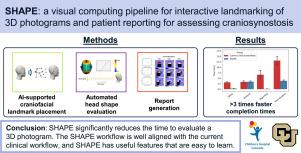
三维摄影测量是一种经济有效的非侵入性成像方式,无需使用电离辐射或镇静剂。因此,它在儿科具有特殊的价值,可用于支持颅面发育病症的诊断和纵向研究,例如颅骨发育不全--一条或多条颅缝过早融合,导致局部颅骨生长受限和颅骨畸形。三维摄影测量分析需要识别颅面地标,以分割头部表面并计算量化异常的指标。遗憾的是,商业三维摄影测量软件需要大量的人工放置地标,既费时又容易出错。我们设计并实施了 SHAPE--头形分析和儿科评估系统。该系统将我们之前开发的自动标记方法集成到视觉计算管道中,以评估患者的三维照片,同时允许手动确认和校正。它还能自动计算量化颅面畸形的高级指标,并自动创建可上传到患者电子健康记录的报告。我们与一名专业临床摄影师进行了用户研究,将 SHAPE 与现有的临床工作流程进行比较。我们发现,SHAPE 评估颅面三维照片的速度比现有临床工作流程快三倍多(3.85±0.99 分钟 vs. 13.07±5.29 分钟,p<0.001)。我们的定性研究结果表明,SHAPE 工作流程与现有的临床工作流程非常吻合,而且 SHAPE 功能实用、易于学习。
本文章由计算机程序翻译,如有差异,请以英文原文为准。

SHAPE: A visual computing pipeline for interactive landmarking of 3D photograms and patient reporting for assessing craniosynostosis

SHAPE: A visual computing pipeline for interactive landmarking of 3D photograms and patient reporting for assessing craniosynostosis
3D photogrammetry is a cost-effective, non-invasive imaging modality that does not require the use of ionizing radiation or sedation. Therefore, it is specifically valuable in pediatrics and is used to support the diagnosis and longitudinal study of craniofacial developmental pathologies such as craniosynostosis — the premature fusion of one or more cranial sutures resulting in local cranial growth restrictions and cranial malformations. Analysis of 3D photogrammetry requires the identification of craniofacial landmarks to segment the head surface and compute metrics to quantify anomalies. Unfortunately, commercial 3D photogrammetry software requires intensive manual landmark placements, which is time-consuming and prone to errors. We designed and implemented SHAPE, a System for Head-shape Analysis and Pediatric Evaluation. It integrates our previously developed automated landmarking method in a visual computing pipeline to evaluate a patient’s 3D photogram while allowing for manual confirmation and correction. It also automatically computes advanced metrics to quantify craniofacial anomalies and automatically creates a report that can be uploaded to the patient’s electronic health record. We conducted a user study with a professional clinical photographer to compare SHAPE to the existing clinical workflow. We found that SHAPE allows for the evaluation of a craniofacial 3D photogram more than three times faster than the current clinical workflow (3.85±0.99 vs. 13.07±5.29 minutes, p<0.001). Our qualitative study findings indicate that the SHAPE workflow is well aligned with the existing clinical workflow and that SHAPE has useful features and is easy to learn.
求助全文
通过发布文献求助,成功后即可免费获取论文全文。 去求助
来源期刊
Computers & Graphics-Uk
Computers & Graphics-Uk 工程技术-计算机:软件工程
CiteScore
5.30
自引率
12.00%
发文量
173
审稿时长
38 days
期刊介绍: Computers & Graphics is dedicated to disseminate information on research and applications of computer graphics (CG) techniques. The journal encourages articles on: 1. Research and applications of interactive computer graphics. We are particularly interested in novel interaction techniques and applications of CG to problem domains. 2. State-of-the-art papers on late-breaking, cutting-edge research on CG. 3. Information on innovative uses of graphics principles and technologies. 4. Tutorial papers on both teaching CG principles and innovative uses of CG in education.
×
引用
GB/T 7714-2015
复制
MLA
复制
APA
复制
导出至
BibTeX EndNote RefMan NoteFirst NoteExpress
×
提示
您的信息不完整,为了账户安全,请先补充。
现在去补充
×
提示
您因"违规操作"
具体请查看互助需知
我知道了
×
提示
确定
请完成安全验证×
copy
已复制链接
快去分享给好友吧!
我知道了
右上角分享
点击右上角分享
0
联系我们:info@booksci.cn Book学术提供免费学术资源搜索服务,方便国内外学者检索中英文文献。致力于提供最便捷和优质的服务体验。 Copyright © 2023 布克学术 All rights reserved.
京ICP备2023020795号-1
ghs 京公网安备 11010802042870号
Book学术文献互助
Book学术文献互助群
群 号:481959085
Book学术官方微信