{"title":"您的诊断结果是什么?一头利木赞公牛的急性溶血。","authors":"Alexandre Bertin, Thomas Bonnet, Matthias Lambert, Emi Ludemann, Fabien Corbière, Corine Boucraut, Marie-Noëlle Lucas, Catherine Trumel","doi":"10.1111/vcp.13397","DOIUrl":null,"url":null,"abstract":"<p>A 4-year-old Limousin bull was presented for necropsy at the Veterinary School of Toulouse. The bull had been on pasture for 2 months. He was depressed and pyrexic (40.9°C) the day before. He was treated with sulfadimidine (90 mg/kg live weight) and flunixin meglumine (2 mg/kg live weight) but died during the night.</p><p>The necropsy was performed within 12 h of death and revealed cachexia, pale mucous membranes, few superficial abomasal ulcers, few petechiae of the epicardium and endocardium, diffuse orange discoloration of the liver, severe splenomegaly (85 × 25 cm) and dark red urine. Centrifugation of the urine confirmed the pigmenturia and was highly suggestive of hemoglobinuria. A fine-needle aspiration (FNA) of the spleen was performed (Figure 1).</p><p>Samples of spleen (Figure 2A,B), kidney (Figure 3), and liver were fixed in formalin and routinely processed for histopathology. The splenic red pulp was severely congested with numerous pinpoint basophilic inclusions within erythrocytes consistent with piroplasms, and numerous activated macrophages, some of which contained a mixed brown to green dark pigment, positive to Perl's Prussian stain. The white pulp was moderately hyperplastic. The renal cortex was diffusely congested with erythrocytic parasites, and some urinary tubules were necrotic with an accumulation of a cytoplasmic brown-to-green pigment, probably a mix of hemoglobin and biliary pigments. Lesions were consistent with a generalized parasitemia associated with intravascular and extravascular hepatocytic and splenic hemolysis responsible for centrolobular anoxic hepatocytic necrosis and cholestasis, bilirubinuric and hemoglobinuric nephrosis, and diffuse reactive splenitis.</p><p>Real-time PCR with high specificity to the 18S RNA gene (<i>Babesia</i> spp./<i>Theileria</i> spp.) was performed on a spleen sample and was strongly positive. Subsequently, a fragment of 959 bp of the 18S RNA gene was sequenced and revealed a high homology (97.2% on 959 bp and 99.7% on an internal fragment of 452 bp) with several <i>Theileria orientalis</i> isolates.</p><p>The macroscopic lesions identified at necropsy, coupled with the presence of suspected hemoglobinuria upon urine centrifugation, strongly suggested intravascular hemolytic anemia. Piroplasmosis is commonly reported in the area where the bull came from and was, therefore, a primary differential. Other major causes of hemoglobinuria include <i>Clostridium perfringens</i> or <i>Leptospira</i> spp. infections, but these were not investigated further after examination of the FNA.</p><p>Theileriosis is a tick-borne disease that causes losses in the cattle industry. Several species of <i>Theileria</i> can infect bovines, with <i>T. annulata</i> and <i>T. parva</i> being highly pathogenic. Conversely, <i>T. orientalis</i> complex usually causes mild or asymptomatic disease in endemic regions. However, an increasing number of oriental theileriosis outbreak cases have been reported in both beef and dairy cattle.<span><sup>1-3</sup></span></p><p>Clinical signs associated with <i>T. orientalis</i> infection are often non-specific. Anemia, icterus, and hemoglobinuria related to intravascular hemolysis are common features. Clinical disease is more frequently seen in naïve cattle introduced into endemic areas or in animals stressed due to pregnancy, lactation, or changes to environmental conditions.<span><sup>4</sup></span> In our case, the necropsy revealed cachexia, and it was hypothesized that the bull was immunocompromised. In Europe, <i>T. orientalis</i> has been reported in asymptomatic cattle from Hungary, Portugal, Italy, and Serbia and was also found co-infected with <i>Anaplasma bovis</i> in three Croatian cows that died suddenly.<span><sup>1, 5</sup></span> <i>Theileria orientalis</i> has been reported in France since the early 20th century, but no clinical signs have been attributed to it.<span><sup>6</sup></span> Its prevalence, although largely unknown, may be underestimated due to clinical confusion with babesiosis or because of asymptomatic infections.</p><p>In sick cattle with hemolytic anemia, a blood smear is an inexpensive test that can differentiate cattle infected with piroplasmosis and other blood parasites from non-infected cattle. However, as the morphology of piroplasm is highly variable, the smear cannot be used to determine whether it is <i>Theileria</i> spp. or <i>Babesia</i> spp., and is even less reliable for identifying the different species of <i>Theileria</i>.<span><sup>7</sup></span> In this case, while a blood smear was not performed, a high parasitemia would have been expected.</p><p>As a screening test to detect <i>T. orientalis</i> in naïve herds, a blood smear may fail to detect low levels of parasitemia. A negative blood smear must be confirmed by PCR.<span><sup>4</sup></span></p><p>PCR is known to have high sensitivity and specificity and can identify the species and type of <i>Theileria</i>. Here, PCR was performed on the spleen, which has previously been reported to be useful for determining species of <i>Theileria</i> using the RNA gene. The genetic homology was the highest for <i>T. orientalis</i>, but co-infections with other <i>Theileria</i> species present in lower levels might not have been detected by sequencing. Based on the MPSP (major piroplasm surface protein) gene sequencing, 11 genotypes (types 1–8 and types N1-3) have been proposed for the <i>T. orientalis</i> group.<span><sup>8</sup></span> Type 1 (Chitose) and type 2 (Ikeda) have been shown to be pathogenic. Sequencing was not performed on this bull.</p><p>To the authors' knowledge, this is the first published cytologic description of the bovine spleen with oriental theileriosis. This observation may be of interest as an inexpensive post-mortem test for diagnosis of intra-erythrocytic parasites.</p><p>The authors declare that they have no conflict of interest.</p>","PeriodicalId":23593,"journal":{"name":"Veterinary clinical pathology","volume":"54 S1","pages":"S54-S56"},"PeriodicalIF":1.1000,"publicationDate":"2024-10-23","publicationTypes":"Journal Article","fieldsOfStudy":null,"isOpenAccess":false,"openAccessPdf":"https://onlinelibrary.wiley.com/doi/epdf/10.1111/vcp.13397","citationCount":"0","resultStr":"{\"title\":\"What is your diagnosis: Acute hemolysis in a Limousin bull\",\"authors\":\"Alexandre Bertin, Thomas Bonnet, Matthias Lambert, Emi Ludemann, Fabien Corbière, Corine Boucraut, Marie-Noëlle Lucas, Catherine Trumel\",\"doi\":\"10.1111/vcp.13397\",\"DOIUrl\":null,\"url\":null,\"abstract\":\"<p>A 4-year-old Limousin bull was presented for necropsy at the Veterinary School of Toulouse. The bull had been on pasture for 2 months. He was depressed and pyrexic (40.9°C) the day before. He was treated with sulfadimidine (90 mg/kg live weight) and flunixin meglumine (2 mg/kg live weight) but died during the night.</p><p>The necropsy was performed within 12 h of death and revealed cachexia, pale mucous membranes, few superficial abomasal ulcers, few petechiae of the epicardium and endocardium, diffuse orange discoloration of the liver, severe splenomegaly (85 × 25 cm) and dark red urine. Centrifugation of the urine confirmed the pigmenturia and was highly suggestive of hemoglobinuria. A fine-needle aspiration (FNA) of the spleen was performed (Figure 1).</p><p>Samples of spleen (Figure 2A,B), kidney (Figure 3), and liver were fixed in formalin and routinely processed for histopathology. The splenic red pulp was severely congested with numerous pinpoint basophilic inclusions within erythrocytes consistent with piroplasms, and numerous activated macrophages, some of which contained a mixed brown to green dark pigment, positive to Perl's Prussian stain. The white pulp was moderately hyperplastic. The renal cortex was diffusely congested with erythrocytic parasites, and some urinary tubules were necrotic with an accumulation of a cytoplasmic brown-to-green pigment, probably a mix of hemoglobin and biliary pigments. Lesions were consistent with a generalized parasitemia associated with intravascular and extravascular hepatocytic and splenic hemolysis responsible for centrolobular anoxic hepatocytic necrosis and cholestasis, bilirubinuric and hemoglobinuric nephrosis, and diffuse reactive splenitis.</p><p>Real-time PCR with high specificity to the 18S RNA gene (<i>Babesia</i> spp./<i>Theileria</i> spp.) was performed on a spleen sample and was strongly positive. Subsequently, a fragment of 959 bp of the 18S RNA gene was sequenced and revealed a high homology (97.2% on 959 bp and 99.7% on an internal fragment of 452 bp) with several <i>Theileria orientalis</i> isolates.</p><p>The macroscopic lesions identified at necropsy, coupled with the presence of suspected hemoglobinuria upon urine centrifugation, strongly suggested intravascular hemolytic anemia. Piroplasmosis is commonly reported in the area where the bull came from and was, therefore, a primary differential. Other major causes of hemoglobinuria include <i>Clostridium perfringens</i> or <i>Leptospira</i> spp. infections, but these were not investigated further after examination of the FNA.</p><p>Theileriosis is a tick-borne disease that causes losses in the cattle industry. Several species of <i>Theileria</i> can infect bovines, with <i>T. annulata</i> and <i>T. parva</i> being highly pathogenic. Conversely, <i>T. orientalis</i> complex usually causes mild or asymptomatic disease in endemic regions. However, an increasing number of oriental theileriosis outbreak cases have been reported in both beef and dairy cattle.<span><sup>1-3</sup></span></p><p>Clinical signs associated with <i>T. orientalis</i> infection are often non-specific. Anemia, icterus, and hemoglobinuria related to intravascular hemolysis are common features. Clinical disease is more frequently seen in naïve cattle introduced into endemic areas or in animals stressed due to pregnancy, lactation, or changes to environmental conditions.<span><sup>4</sup></span> In our case, the necropsy revealed cachexia, and it was hypothesized that the bull was immunocompromised. In Europe, <i>T. orientalis</i> has been reported in asymptomatic cattle from Hungary, Portugal, Italy, and Serbia and was also found co-infected with <i>Anaplasma bovis</i> in three Croatian cows that died suddenly.<span><sup>1, 5</sup></span> <i>Theileria orientalis</i> has been reported in France since the early 20th century, but no clinical signs have been attributed to it.<span><sup>6</sup></span> Its prevalence, although largely unknown, may be underestimated due to clinical confusion with babesiosis or because of asymptomatic infections.</p><p>In sick cattle with hemolytic anemia, a blood smear is an inexpensive test that can differentiate cattle infected with piroplasmosis and other blood parasites from non-infected cattle. However, as the morphology of piroplasm is highly variable, the smear cannot be used to determine whether it is <i>Theileria</i> spp. or <i>Babesia</i> spp., and is even less reliable for identifying the different species of <i>Theileria</i>.<span><sup>7</sup></span> In this case, while a blood smear was not performed, a high parasitemia would have been expected.</p><p>As a screening test to detect <i>T. orientalis</i> in naïve herds, a blood smear may fail to detect low levels of parasitemia. A negative blood smear must be confirmed by PCR.<span><sup>4</sup></span></p><p>PCR is known to have high sensitivity and specificity and can identify the species and type of <i>Theileria</i>. Here, PCR was performed on the spleen, which has previously been reported to be useful for determining species of <i>Theileria</i> using the RNA gene. The genetic homology was the highest for <i>T. orientalis</i>, but co-infections with other <i>Theileria</i> species present in lower levels might not have been detected by sequencing. Based on the MPSP (major piroplasm surface protein) gene sequencing, 11 genotypes (types 1–8 and types N1-3) have been proposed for the <i>T. orientalis</i> group.<span><sup>8</sup></span> Type 1 (Chitose) and type 2 (Ikeda) have been shown to be pathogenic. Sequencing was not performed on this bull.</p><p>To the authors' knowledge, this is the first published cytologic description of the bovine spleen with oriental theileriosis. This observation may be of interest as an inexpensive post-mortem test for diagnosis of intra-erythrocytic parasites.</p><p>The authors declare that they have no conflict of interest.</p>\",\"PeriodicalId\":23593,\"journal\":{\"name\":\"Veterinary clinical pathology\",\"volume\":\"54 S1\",\"pages\":\"S54-S56\"},\"PeriodicalIF\":1.1000,\"publicationDate\":\"2024-10-23\",\"publicationTypes\":\"Journal Article\",\"fieldsOfStudy\":null,\"isOpenAccess\":false,\"openAccessPdf\":\"https://onlinelibrary.wiley.com/doi/epdf/10.1111/vcp.13397\",\"citationCount\":\"0\",\"resultStr\":null,\"platform\":\"Semanticscholar\",\"paperid\":null,\"PeriodicalName\":\"Veterinary clinical pathology\",\"FirstCategoryId\":\"97\",\"ListUrlMain\":\"https://onlinelibrary.wiley.com/doi/10.1111/vcp.13397\",\"RegionNum\":4,\"RegionCategory\":\"农林科学\",\"ArticlePicture\":[],\"TitleCN\":null,\"AbstractTextCN\":null,\"PMCID\":null,\"EPubDate\":\"\",\"PubModel\":\"\",\"JCR\":\"Q3\",\"JCRName\":\"VETERINARY SCIENCES\",\"Score\":null,\"Total\":0}","platform":"Semanticscholar","paperid":null,"PeriodicalName":"Veterinary clinical pathology","FirstCategoryId":"97","ListUrlMain":"https://onlinelibrary.wiley.com/doi/10.1111/vcp.13397","RegionNum":4,"RegionCategory":"农林科学","ArticlePicture":[],"TitleCN":null,"AbstractTextCN":null,"PMCID":null,"EPubDate":"","PubModel":"","JCR":"Q3","JCRName":"VETERINARY SCIENCES","Score":null,"Total":0}
What is your diagnosis: Acute hemolysis in a Limousin bull
A 4-year-old Limousin bull was presented for necropsy at the Veterinary School of Toulouse. The bull had been on pasture for 2 months. He was depressed and pyrexic (40.9°C) the day before. He was treated with sulfadimidine (90 mg/kg live weight) and flunixin meglumine (2 mg/kg live weight) but died during the night.
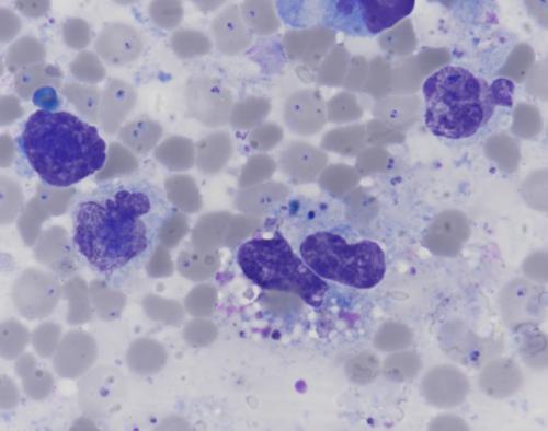
The necropsy was performed within 12 h of death and revealed cachexia, pale mucous membranes, few superficial abomasal ulcers, few petechiae of the epicardium and endocardium, diffuse orange discoloration of the liver, severe splenomegaly (85 × 25 cm) and dark red urine. Centrifugation of the urine confirmed the pigmenturia and was highly suggestive of hemoglobinuria. A fine-needle aspiration (FNA) of the spleen was performed (Figure 1).
Samples of spleen (Figure 2A,B), kidney (Figure 3), and liver were fixed in formalin and routinely processed for histopathology. The splenic red pulp was severely congested with numerous pinpoint basophilic inclusions within erythrocytes consistent with piroplasms, and numerous activated macrophages, some of which contained a mixed brown to green dark pigment, positive to Perl's Prussian stain. The white pulp was moderately hyperplastic. The renal cortex was diffusely congested with erythrocytic parasites, and some urinary tubules were necrotic with an accumulation of a cytoplasmic brown-to-green pigment, probably a mix of hemoglobin and biliary pigments. Lesions were consistent with a generalized parasitemia associated with intravascular and extravascular hepatocytic and splenic hemolysis responsible for centrolobular anoxic hepatocytic necrosis and cholestasis, bilirubinuric and hemoglobinuric nephrosis, and diffuse reactive splenitis.
Real-time PCR with high specificity to the 18S RNA gene (Babesia spp./Theileria spp.) was performed on a spleen sample and was strongly positive. Subsequently, a fragment of 959 bp of the 18S RNA gene was sequenced and revealed a high homology (97.2% on 959 bp and 99.7% on an internal fragment of 452 bp) with several Theileria orientalis isolates.
The macroscopic lesions identified at necropsy, coupled with the presence of suspected hemoglobinuria upon urine centrifugation, strongly suggested intravascular hemolytic anemia. Piroplasmosis is commonly reported in the area where the bull came from and was, therefore, a primary differential. Other major causes of hemoglobinuria include Clostridium perfringens or Leptospira spp. infections, but these were not investigated further after examination of the FNA.
Theileriosis is a tick-borne disease that causes losses in the cattle industry. Several species of Theileria can infect bovines, with T. annulata and T. parva being highly pathogenic. Conversely, T. orientalis complex usually causes mild or asymptomatic disease in endemic regions. However, an increasing number of oriental theileriosis outbreak cases have been reported in both beef and dairy cattle.1-3
Clinical signs associated with T. orientalis infection are often non-specific. Anemia, icterus, and hemoglobinuria related to intravascular hemolysis are common features. Clinical disease is more frequently seen in naïve cattle introduced into endemic areas or in animals stressed due to pregnancy, lactation, or changes to environmental conditions.4 In our case, the necropsy revealed cachexia, and it was hypothesized that the bull was immunocompromised. In Europe, T. orientalis has been reported in asymptomatic cattle from Hungary, Portugal, Italy, and Serbia and was also found co-infected with Anaplasma bovis in three Croatian cows that died suddenly.1, 5Theileria orientalis has been reported in France since the early 20th century, but no clinical signs have been attributed to it.6 Its prevalence, although largely unknown, may be underestimated due to clinical confusion with babesiosis or because of asymptomatic infections.
In sick cattle with hemolytic anemia, a blood smear is an inexpensive test that can differentiate cattle infected with piroplasmosis and other blood parasites from non-infected cattle. However, as the morphology of piroplasm is highly variable, the smear cannot be used to determine whether it is Theileria spp. or Babesia spp., and is even less reliable for identifying the different species of Theileria.7 In this case, while a blood smear was not performed, a high parasitemia would have been expected.
As a screening test to detect T. orientalis in naïve herds, a blood smear may fail to detect low levels of parasitemia. A negative blood smear must be confirmed by PCR.4
PCR is known to have high sensitivity and specificity and can identify the species and type of Theileria. Here, PCR was performed on the spleen, which has previously been reported to be useful for determining species of Theileria using the RNA gene. The genetic homology was the highest for T. orientalis, but co-infections with other Theileria species present in lower levels might not have been detected by sequencing. Based on the MPSP (major piroplasm surface protein) gene sequencing, 11 genotypes (types 1–8 and types N1-3) have been proposed for the T. orientalis group.8 Type 1 (Chitose) and type 2 (Ikeda) have been shown to be pathogenic. Sequencing was not performed on this bull.
To the authors' knowledge, this is the first published cytologic description of the bovine spleen with oriental theileriosis. This observation may be of interest as an inexpensive post-mortem test for diagnosis of intra-erythrocytic parasites.
The authors declare that they have no conflict of interest.
期刊介绍:
Veterinary Clinical Pathology is the official journal of the American Society for Veterinary Clinical Pathology (ASVCP) and the European Society of Veterinary Clinical Pathology (ESVCP). The journal''s mission is to provide an international forum for communication and discussion of scientific investigations and new developments that advance the art and science of laboratory diagnosis in animals. Veterinary Clinical Pathology welcomes original experimental research and clinical contributions involving domestic, laboratory, avian, and wildlife species in the areas of hematology, hemostasis, immunopathology, clinical chemistry, cytopathology, surgical pathology, toxicology, endocrinology, laboratory and analytical techniques, instrumentation, quality assurance, and clinical pathology education.
 求助内容:
求助内容: 应助结果提醒方式:


