Digvijay D Nalawade, Pratik Satyajit Wadhokar, Ajitkumar Krishna Jadhav, Vivek V Manade
{"title":"一例永久性起搏器植入术中模仿右心房血栓的突出嵴端--二维和三维经食道超声心动图的作用。","authors":"Digvijay D Nalawade, Pratik Satyajit Wadhokar, Ajitkumar Krishna Jadhav, Vivek V Manade","doi":"10.4103/jcecho.jcecho_2_23","DOIUrl":null,"url":null,"abstract":"<p><p>Crista terminalis is a crescent-shaped fibromuscular ridge in the posterolateral wall of the right atrium (RA) which separates the smooth posterior region of RA from a more muscular anterior region. When prominent, it frequently mimics RA thrombus, vegetation, or tumors such as myxoma. Differentiation of such anatomical structural variations from other masses is vital to minimize misdiagnosis and avoid disease-related apprehension. Different diagnostic modalities may be needed which have their own imaging characteristics as well as limitations. Our case emphasizes the differentiating features of prominent crista terminalis using two-dimensional and three-dimensional transesophageal echocardiography.</p>","PeriodicalId":15191,"journal":{"name":"Journal of Cardiovascular Echography","volume":"34 3","pages":"149-151"},"PeriodicalIF":0.7000,"publicationDate":"2024-07-01","publicationTypes":"Journal Article","fieldsOfStudy":null,"isOpenAccess":false,"openAccessPdf":"https://www.ncbi.nlm.nih.gov/pmc/articles/PMC11495316/pdf/","citationCount":"0","resultStr":"{\"title\":\"Prominent Crista Terminalis Mimicking Right Atrial Thrombus in a Case of Permanent Pacemaker Implantation - Role of Two- and Three-Dimensional Transesophageal Echocardiography.\",\"authors\":\"Digvijay D Nalawade, Pratik Satyajit Wadhokar, Ajitkumar Krishna Jadhav, Vivek V Manade\",\"doi\":\"10.4103/jcecho.jcecho_2_23\",\"DOIUrl\":null,\"url\":null,\"abstract\":\"<p><p>Crista terminalis is a crescent-shaped fibromuscular ridge in the posterolateral wall of the right atrium (RA) which separates the smooth posterior region of RA from a more muscular anterior region. When prominent, it frequently mimics RA thrombus, vegetation, or tumors such as myxoma. Differentiation of such anatomical structural variations from other masses is vital to minimize misdiagnosis and avoid disease-related apprehension. Different diagnostic modalities may be needed which have their own imaging characteristics as well as limitations. Our case emphasizes the differentiating features of prominent crista terminalis using two-dimensional and three-dimensional transesophageal echocardiography.</p>\",\"PeriodicalId\":15191,\"journal\":{\"name\":\"Journal of Cardiovascular Echography\",\"volume\":\"34 3\",\"pages\":\"149-151\"},\"PeriodicalIF\":0.7000,\"publicationDate\":\"2024-07-01\",\"publicationTypes\":\"Journal Article\",\"fieldsOfStudy\":null,\"isOpenAccess\":false,\"openAccessPdf\":\"https://www.ncbi.nlm.nih.gov/pmc/articles/PMC11495316/pdf/\",\"citationCount\":\"0\",\"resultStr\":null,\"platform\":\"Semanticscholar\",\"paperid\":null,\"PeriodicalName\":\"Journal of Cardiovascular Echography\",\"FirstCategoryId\":\"1085\",\"ListUrlMain\":\"https://doi.org/10.4103/jcecho.jcecho_2_23\",\"RegionNum\":0,\"RegionCategory\":null,\"ArticlePicture\":[],\"TitleCN\":null,\"AbstractTextCN\":null,\"PMCID\":null,\"EPubDate\":\"2024/9/21 0:00:00\",\"PubModel\":\"Epub\",\"JCR\":\"Q4\",\"JCRName\":\"CARDIAC & CARDIOVASCULAR SYSTEMS\",\"Score\":null,\"Total\":0}","platform":"Semanticscholar","paperid":null,"PeriodicalName":"Journal of Cardiovascular Echography","FirstCategoryId":"1085","ListUrlMain":"https://doi.org/10.4103/jcecho.jcecho_2_23","RegionNum":0,"RegionCategory":null,"ArticlePicture":[],"TitleCN":null,"AbstractTextCN":null,"PMCID":null,"EPubDate":"2024/9/21 0:00:00","PubModel":"Epub","JCR":"Q4","JCRName":"CARDIAC & CARDIOVASCULAR SYSTEMS","Score":null,"Total":0}
引用次数: 0
摘要
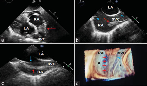
终末嵴是右心房(RA)后外侧壁上的一个新月形纤维肌脊,它将右心房光滑的后部区域与肌肉发达的前部区域分隔开来。当突出时,它经常模仿 RA 血栓、植被或肿瘤(如肌瘤)。将这种解剖结构上的变异与其他肿块区分开来对于减少误诊和避免与疾病相关的忧虑至关重要。可能需要采用不同的诊断方法,这些方法都有各自的成像特点和局限性。我们的病例强调了使用二维和三维经食道超声心动图对突出的嵴突进行鉴别的特点。
Prominent Crista Terminalis Mimicking Right Atrial Thrombus in a Case of Permanent Pacemaker Implantation - Role of Two- and Three-Dimensional Transesophageal Echocardiography.
Crista terminalis is a crescent-shaped fibromuscular ridge in the posterolateral wall of the right atrium (RA) which separates the smooth posterior region of RA from a more muscular anterior region. When prominent, it frequently mimics RA thrombus, vegetation, or tumors such as myxoma. Differentiation of such anatomical structural variations from other masses is vital to minimize misdiagnosis and avoid disease-related apprehension. Different diagnostic modalities may be needed which have their own imaging characteristics as well as limitations. Our case emphasizes the differentiating features of prominent crista terminalis using two-dimensional and three-dimensional transesophageal echocardiography.