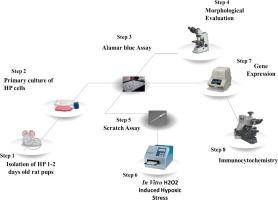
奎宁酸有助于神经发生:以海马中的关键角色 Notch 通路为目标

IF 2.7 4区 医学 Q3 NEUROSCIENCES
Maryam Niaz , Kanwal Iftikhar , Maha Shahid , Shaheen Faizi , Shabana Usman Simjee
{"title":"奎宁酸有助于神经发生:以海马中的关键角色 Notch 通路为目标","authors":"Maryam Niaz ,&nbsp;Kanwal Iftikhar ,&nbsp;Maha Shahid ,&nbsp;Shaheen Faizi ,&nbsp;Shabana Usman Simjee","doi":"10.1016/j.brainres.2024.149291","DOIUrl":null,"url":null,"abstract":"<div><div>Coordinated proliferation and differentiation of neural stem cells (NSCs) results in continuous neurogenesis. The present study provides novel insights into the Notch intracellular signaling in neuronal cell proliferation, maintenance, migration, and differentiation regulated by naturally based Quinic acid (QA) in primary hippocampal cell culture. Further, this study might help in the discovery and development of lead molecules that can overcome the challenges in the treatment of neurodegenerative diseases. The growth supporting effect of QA was studied using Alamar Blue assay. The migratory potential of QA was evaluated using scratch assay. The <em>in vitro</em> H<sub>2</sub>O<sub>2</sub>-induced oxidative stress model was used to upregulate neuronal survival after QA treatment. The RT-qPCR and immunocytochemical analysis were performed for selected markers of Notch signaling to determine the proliferation, differentiation, and maintenance of NSCs at gene and molecular levels. The Mash1 and Ngn2 are the upstream proneural genes of the Notch pathway which were included to evaluate the differentiation of NSCs into mature neurons after treatment with QA.</div><div>Furthermore, regarding the role of QA in maintaining the pool of NPCs, we used Notch1 and Hes1 markers for proliferation analysis. Also, secondary neuronal markers i.e. Pax6, PCNA<strong>,</strong> and Mcm2 were included in this study and their gene expression analysis was analyzed following treatment with QA. Based on the study’s results, we suggest that naturally based QA can promote the growth and differentiation of neonatal NSCs residing in hippocampal regions into neuronal lineage. Therefore, we propose that the neurogenic potential of QA can be employed to prevent and treat neurodegenerative diseases.</div></div>","PeriodicalId":9083,"journal":{"name":"Brain Research","volume":"1846 ","pages":"Article 149291"},"PeriodicalIF":2.7000,"publicationDate":"2024-10-22","publicationTypes":"Journal Article","fieldsOfStudy":null,"isOpenAccess":false,"openAccessPdf":"","citationCount":"0","resultStr":"{\"title\":\"Quinic acid contributes to neurogenesis: Targeting Notch pathway a key player in hippocampus\",\"authors\":\"Maryam Niaz ,&nbsp;Kanwal Iftikhar ,&nbsp;Maha Shahid ,&nbsp;Shaheen Faizi ,&nbsp;Shabana Usman Simjee\",\"doi\":\"10.1016/j.brainres.2024.149291\",\"DOIUrl\":null,\"url\":null,\"abstract\":\"<div><div>Coordinated proliferation and differentiation of neural stem cells (NSCs) results in continuous neurogenesis. The present study provides novel insights into the Notch intracellular signaling in neuronal cell proliferation, maintenance, migration, and differentiation regulated by naturally based Quinic acid (QA) in primary hippocampal cell culture. Further, this study might help in the discovery and development of lead molecules that can overcome the challenges in the treatment of neurodegenerative diseases. The growth supporting effect of QA was studied using Alamar Blue assay. The migratory potential of QA was evaluated using scratch assay. The <em>in vitro</em> H<sub>2</sub>O<sub>2</sub>-induced oxidative stress model was used to upregulate neuronal survival after QA treatment. The RT-qPCR and immunocytochemical analysis were performed for selected markers of Notch signaling to determine the proliferation, differentiation, and maintenance of NSCs at gene and molecular levels. The Mash1 and Ngn2 are the upstream proneural genes of the Notch pathway which were included to evaluate the differentiation of NSCs into mature neurons after treatment with QA.</div><div>Furthermore, regarding the role of QA in maintaining the pool of NPCs, we used Notch1 and Hes1 markers for proliferation analysis. Also, secondary neuronal markers i.e. Pax6, PCNA<strong>,</strong> and Mcm2 were included in this study and their gene expression analysis was analyzed following treatment with QA. Based on the study’s results, we suggest that naturally based QA can promote the growth and differentiation of neonatal NSCs residing in hippocampal regions into neuronal lineage. Therefore, we propose that the neurogenic potential of QA can be employed to prevent and treat neurodegenerative diseases.</div></div>\",\"PeriodicalId\":9083,\"journal\":{\"name\":\"Brain Research\",\"volume\":\"1846 \",\"pages\":\"Article 149291\"},\"PeriodicalIF\":2.7000,\"publicationDate\":\"2024-10-22\",\"publicationTypes\":\"Journal Article\",\"fieldsOfStudy\":null,\"isOpenAccess\":false,\"openAccessPdf\":\"\",\"citationCount\":\"0\",\"resultStr\":null,\"platform\":\"Semanticscholar\",\"paperid\":null,\"PeriodicalName\":\"Brain Research\",\"FirstCategoryId\":\"3\",\"ListUrlMain\":\"https://www.sciencedirect.com/science/article/pii/S0006899324005456\",\"RegionNum\":4,\"RegionCategory\":\"医学\",\"ArticlePicture\":[],\"TitleCN\":null,\"AbstractTextCN\":null,\"PMCID\":null,\"EPubDate\":\"\",\"PubModel\":\"\",\"JCR\":\"Q3\",\"JCRName\":\"NEUROSCIENCES\",\"Score\":null,\"Total\":0}","platform":"Semanticscholar","paperid":null,"PeriodicalName":"Brain Research","FirstCategoryId":"3","ListUrlMain":"https://www.sciencedirect.com/science/article/pii/S0006899324005456","RegionNum":4,"RegionCategory":"医学","ArticlePicture":[],"TitleCN":null,"AbstractTextCN":null,"PMCID":null,"EPubDate":"","PubModel":"","JCR":"Q3","JCRName":"NEUROSCIENCES","Score":null,"Total":0}
引用次数: 0

摘要

神经干细胞(NSCs)的协调增殖和分化可导致持续的神经发生。本研究提供了关于天然奎尼酸(QA)在原代海马细胞培养中调控神经细胞增殖、维持、迁移和分化的细胞内Notch信号的新见解。此外,这项研究可能有助于发现和开发先导分子,从而克服治疗神经退行性疾病的难题。使用阿拉玛蓝检测法研究了 QA 的生长支持效应。划痕试验评估了 QA 的迁移潜力。体外 H2O2 诱导的氧化应激模型用于上调 QA 处理后神经元的存活率。对选定的 Notch 信号转导标记物进行 RT-qPCR 和免疫细胞化学分析,以确定 NSCs 在基因和分子水平上的增殖、分化和维持。其中,Mash1和Ngn2是Notch通路的上游朊病毒基因,它们被用于评估经QA处理后NSCs向成熟神经元的分化。此外,关于 QA 在维持 NPCs 池中的作用,我们使用 Notch1 和 Hes1 标记进行增殖分析。本研究还纳入了次要神经元标记,即 Pax6、PCNA 和 Mcm2,并分析了它们在 QA 处理后的基因表达分析。根据研究结果,我们认为天然 QA 可促进海马区新生儿神经干细胞的生长和分化,使其向神经元系分化。因此,我们建议利用 QA 的神经源潜力来预防和治疗神经退行性疾病。
本文章由计算机程序翻译,如有差异,请以英文原文为准。

Quinic acid contributes to neurogenesis: Targeting Notch pathway a key player in hippocampus

Quinic acid contributes to neurogenesis: Targeting Notch pathway a key player in hippocampus
Coordinated proliferation and differentiation of neural stem cells (NSCs) results in continuous neurogenesis. The present study provides novel insights into the Notch intracellular signaling in neuronal cell proliferation, maintenance, migration, and differentiation regulated by naturally based Quinic acid (QA) in primary hippocampal cell culture. Further, this study might help in the discovery and development of lead molecules that can overcome the challenges in the treatment of neurodegenerative diseases. The growth supporting effect of QA was studied using Alamar Blue assay. The migratory potential of QA was evaluated using scratch assay. The in vitro H2O2-induced oxidative stress model was used to upregulate neuronal survival after QA treatment. The RT-qPCR and immunocytochemical analysis were performed for selected markers of Notch signaling to determine the proliferation, differentiation, and maintenance of NSCs at gene and molecular levels. The Mash1 and Ngn2 are the upstream proneural genes of the Notch pathway which were included to evaluate the differentiation of NSCs into mature neurons after treatment with QA.
Furthermore, regarding the role of QA in maintaining the pool of NPCs, we used Notch1 and Hes1 markers for proliferation analysis. Also, secondary neuronal markers i.e. Pax6, PCNA, and Mcm2 were included in this study and their gene expression analysis was analyzed following treatment with QA. Based on the study’s results, we suggest that naturally based QA can promote the growth and differentiation of neonatal NSCs residing in hippocampal regions into neuronal lineage. Therefore, we propose that the neurogenic potential of QA can be employed to prevent and treat neurodegenerative diseases.
求助全文
通过发布文献求助,成功后即可免费获取论文全文。 去求助
来源期刊
Brain Research
Brain Research 医学-神经科学
CiteScore
5.90
自引率
3.40%
发文量
268
审稿时长
47 days
期刊介绍: An international multidisciplinary journal devoted to fundamental research in the brain sciences. Brain Research publishes papers reporting interdisciplinary investigations of nervous system structure and function that are of general interest to the international community of neuroscientists. As is evident from the journals name, its scope is broad, ranging from cellular and molecular studies through systems neuroscience, cognition and disease. Invited reviews are also published; suggestions for and inquiries about potential reviews are welcomed. With the appearance of the final issue of the 2011 subscription, Vol. 67/1-2 (24 June 2011), Brain Research Reviews has ceased publication as a distinct journal separate from Brain Research. Review articles accepted for Brain Research are now published in that journal.
×
引用
GB/T 7714-2015
复制
MLA
复制
APA
复制
导出至
BibTeX EndNote RefMan NoteFirst NoteExpress
×
提示
您的信息不完整,为了账户安全,请先补充。
现在去补充
×
提示
您因"违规操作"
具体请查看互助需知
我知道了
×
提示
确定
请完成安全验证×
copy
已复制链接
快去分享给好友吧!
我知道了
右上角分享
点击右上角分享
0
联系我们:info@booksci.cn Book学术提供免费学术资源搜索服务,方便国内外学者检索中英文文献。致力于提供最便捷和优质的服务体验。 Copyright © 2023 布克学术 All rights reserved.
京ICP备2023020795号-1
ghs 京公网安备 11010802042870号
Book学术文献互助
Book学术文献互助群
群 号:481959085
Book学术官方微信