{"title":"一枚硬币的两面:免疫功能正常的年轻人患上的嗜酸性粒细胞性食管炎和疱疹性食管炎","authors":"Sandra Correia, Tiago Pereira Guedes, Maria Mexia Leitão, Isabel Pedroto, Sílvia Barrias","doi":"10.17235/reed.2024.10839/2024","DOIUrl":null,"url":null,"abstract":"<p><p>Dear Editor, We report a case of a 30-year-old woman with an 8-year diagnosis of eosinophilic esophagitis (EoE) treated with swallowed fluticasone propionate throughout this period. She presented to the emergency room with a two-day history of severe odynophagia, aphagia, retrosternal pain, and fever. The patient was febrile and hemodynamically stable, with no visible oropharyngeal lesions. She presented with elevated C-reactive protein (37 mg/L). An esophagogastroduodenoscopy was performed, which revealed white plaque-like lesions with \"volcano-like\" shallow ulcerations with raised edges on the distal esophagus (Figure 1aandb). Multiple biopsies were taken from both the center and edges of the lesions. The patient was empirically started on intravenous fluconazole due to the suspicion of candida esophagitis. However, the patient's symptoms worsened over the next two days, and acyclovir at a dose of 5 mg/kg was started. The initial work-up showed a positive titer for Herpes Simplex Virus (HSV)-2 IgM (1.6 U/L) and a negative titer for IgG (2.24 U/L), as well as a negative serological study for HSV-1, cytomegalovirus, and human immunodeficiency virus (HIV). Histological examination revealed multinucleated giant cells with nuclear molding and chromatin margination and cells with \"ground glass\" nuclei, along with typical Cowdry type A intranuclear inclusion bodies and immunohistochemical staining for HSV type 2, confirmed the diagnosis of herpetic esophagitis (Figure 1candd). The patient experienced rapid improvement and was discharged on oral acyclovir therapy at 400 mg/day, completing a total of 14 days of treatment with a total resolution of symptoms.</p>","PeriodicalId":21342,"journal":{"name":"Revista Espanola De Enfermedades Digestivas","volume":" ","pages":""},"PeriodicalIF":2.7000,"publicationDate":"2024-10-18","publicationTypes":"Journal Article","fieldsOfStudy":null,"isOpenAccess":false,"openAccessPdf":"","citationCount":"0","resultStr":"{\"title\":\"Two Sides of the Same Coin: Eosinophilic and Herpetic Esophagitis in an Immunocompetent Young Adult.\",\"authors\":\"Sandra Correia, Tiago Pereira Guedes, Maria Mexia Leitão, Isabel Pedroto, Sílvia Barrias\",\"doi\":\"10.17235/reed.2024.10839/2024\",\"DOIUrl\":null,\"url\":null,\"abstract\":\"<p><p>Dear Editor, We report a case of a 30-year-old woman with an 8-year diagnosis of eosinophilic esophagitis (EoE) treated with swallowed fluticasone propionate throughout this period. She presented to the emergency room with a two-day history of severe odynophagia, aphagia, retrosternal pain, and fever. The patient was febrile and hemodynamically stable, with no visible oropharyngeal lesions. She presented with elevated C-reactive protein (37 mg/L). An esophagogastroduodenoscopy was performed, which revealed white plaque-like lesions with \\\"volcano-like\\\" shallow ulcerations with raised edges on the distal esophagus (Figure 1aandb). Multiple biopsies were taken from both the center and edges of the lesions. The patient was empirically started on intravenous fluconazole due to the suspicion of candida esophagitis. However, the patient's symptoms worsened over the next two days, and acyclovir at a dose of 5 mg/kg was started. The initial work-up showed a positive titer for Herpes Simplex Virus (HSV)-2 IgM (1.6 U/L) and a negative titer for IgG (2.24 U/L), as well as a negative serological study for HSV-1, cytomegalovirus, and human immunodeficiency virus (HIV). Histological examination revealed multinucleated giant cells with nuclear molding and chromatin margination and cells with \\\"ground glass\\\" nuclei, along with typical Cowdry type A intranuclear inclusion bodies and immunohistochemical staining for HSV type 2, confirmed the diagnosis of herpetic esophagitis (Figure 1candd). The patient experienced rapid improvement and was discharged on oral acyclovir therapy at 400 mg/day, completing a total of 14 days of treatment with a total resolution of symptoms.</p>\",\"PeriodicalId\":21342,\"journal\":{\"name\":\"Revista Espanola De Enfermedades Digestivas\",\"volume\":\" \",\"pages\":\"\"},\"PeriodicalIF\":2.7000,\"publicationDate\":\"2024-10-18\",\"publicationTypes\":\"Journal Article\",\"fieldsOfStudy\":null,\"isOpenAccess\":false,\"openAccessPdf\":\"\",\"citationCount\":\"0\",\"resultStr\":null,\"platform\":\"Semanticscholar\",\"paperid\":null,\"PeriodicalName\":\"Revista Espanola De Enfermedades Digestivas\",\"FirstCategoryId\":\"3\",\"ListUrlMain\":\"https://doi.org/10.17235/reed.2024.10839/2024\",\"RegionNum\":4,\"RegionCategory\":\"医学\",\"ArticlePicture\":[],\"TitleCN\":null,\"AbstractTextCN\":null,\"PMCID\":null,\"EPubDate\":\"\",\"PubModel\":\"\",\"JCR\":\"Q2\",\"JCRName\":\"GASTROENTEROLOGY & HEPATOLOGY\",\"Score\":null,\"Total\":0}","platform":"Semanticscholar","paperid":null,"PeriodicalName":"Revista Espanola De Enfermedades Digestivas","FirstCategoryId":"3","ListUrlMain":"https://doi.org/10.17235/reed.2024.10839/2024","RegionNum":4,"RegionCategory":"医学","ArticlePicture":[],"TitleCN":null,"AbstractTextCN":null,"PMCID":null,"EPubDate":"","PubModel":"","JCR":"Q2","JCRName":"GASTROENTEROLOGY & HEPATOLOGY","Score":null,"Total":0}
Two Sides of the Same Coin: Eosinophilic and Herpetic Esophagitis in an Immunocompetent Young Adult.
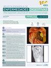
Dear Editor, We report a case of a 30-year-old woman with an 8-year diagnosis of eosinophilic esophagitis (EoE) treated with swallowed fluticasone propionate throughout this period. She presented to the emergency room with a two-day history of severe odynophagia, aphagia, retrosternal pain, and fever. The patient was febrile and hemodynamically stable, with no visible oropharyngeal lesions. She presented with elevated C-reactive protein (37 mg/L). An esophagogastroduodenoscopy was performed, which revealed white plaque-like lesions with "volcano-like" shallow ulcerations with raised edges on the distal esophagus (Figure 1aandb). Multiple biopsies were taken from both the center and edges of the lesions. The patient was empirically started on intravenous fluconazole due to the suspicion of candida esophagitis. However, the patient's symptoms worsened over the next two days, and acyclovir at a dose of 5 mg/kg was started. The initial work-up showed a positive titer for Herpes Simplex Virus (HSV)-2 IgM (1.6 U/L) and a negative titer for IgG (2.24 U/L), as well as a negative serological study for HSV-1, cytomegalovirus, and human immunodeficiency virus (HIV). Histological examination revealed multinucleated giant cells with nuclear molding and chromatin margination and cells with "ground glass" nuclei, along with typical Cowdry type A intranuclear inclusion bodies and immunohistochemical staining for HSV type 2, confirmed the diagnosis of herpetic esophagitis (Figure 1candd). The patient experienced rapid improvement and was discharged on oral acyclovir therapy at 400 mg/day, completing a total of 14 days of treatment with a total resolution of symptoms.
期刊介绍:
La Revista Española de Enfermedades Digestivas, Órgano Oficial de la Sociedad Española de Patología Digestiva (SEPD), Sociedad Española de Endoscopia Digestiva (SEED) y Asociación Española de Ecografía Digestiva (AEED), publica artículos originales, editoriales, revisiones, casos clínicos, cartas al director, imágenes en patología digestiva, y otros artículos especiales sobre todos los aspectos relativos a las enfermedades digestivas.
 求助内容:
求助内容: 应助结果提醒方式:


