{"title":"上皮细胞分裂的乳腺内标记。","authors":"Maia N Machiela, Russell C Hovey","doi":"10.1007/s10911-024-09570-4","DOIUrl":null,"url":null,"abstract":"<p><p>Thymidine analogs such as ethynyl deoxyuridine (EdU) or bromodeoxyuridine (BrdU) can be used to label mitosis of mammary epithelial cells (MEC) and to quantify their proliferation. However, labeling cells in larger animals requires considerable amounts of chemical that can be costly and hazardous. We developed a strategy to infuse EdU into the mammary glands of ewes to directly label mitotic MEC. First, each udder half of nulliparous ewes (n = 2) received an intramammary infusion of one of four different concentrations of EdU (0, 0.1, 1.0 or 10 mM) which was compared to BrdU IV (5 mg/kg) 24 h later. Tissues were analyzed by immunofluorescent histochemistry to detect EdU, BrdU, and total MEC. Of the EdU doses tested, 10 mM EdU yielded the greatest labeling index, while a proportion of MEC were labeled by both EdU and BrdU. We next sought to establish whether intramammary labeling could detect the induction of mitosis after exposure to exogenous estrogen and progesterone (E + P). We first infused EdU (10 mM) into the right udder half of ewes (n = 6) at t 0, followed by thymidine (100 mM) 24 h later to prevent further labeling. Three ewes were then administered E + P for 5 d, while n = 3 ewes served as controls. On d 5, EdU was infused into the left udder half of all mammary glands alongside BrdU IV (5 mg/kg). By the time of necropsy 24 h later an average MEC labeling index of 2.9% resulted from EdU delivered at t 0. In the left half of the udder on d 5, CON glands had a final EdU labeling index of 3.4% while glands exposed to E + P had a labeling index of 4.6% (p = 0.05). The corresponding degree of labeling with BrdU was 5.6% in CON glands, and 12% following E + P (p < 0.001). Our findings reveal that intramammary labeling is an efficient and cost-effective method for single- and dual-labeling of cell division in the mammary glands.</p>","PeriodicalId":16413,"journal":{"name":"Journal of Mammary Gland Biology and Neoplasia","volume":"29 1","pages":"17"},"PeriodicalIF":3.6000,"publicationDate":"2024-10-16","publicationTypes":"Journal Article","fieldsOfStudy":null,"isOpenAccess":false,"openAccessPdf":"https://www.ncbi.nlm.nih.gov/pmc/articles/PMC11485144/pdf/","citationCount":"0","resultStr":"{\"title\":\"Intramammary Labeling of Epithelial Cell Division.\",\"authors\":\"Maia N Machiela, Russell C Hovey\",\"doi\":\"10.1007/s10911-024-09570-4\",\"DOIUrl\":null,\"url\":null,\"abstract\":\"<p><p>Thymidine analogs such as ethynyl deoxyuridine (EdU) or bromodeoxyuridine (BrdU) can be used to label mitosis of mammary epithelial cells (MEC) and to quantify their proliferation. However, labeling cells in larger animals requires considerable amounts of chemical that can be costly and hazardous. We developed a strategy to infuse EdU into the mammary glands of ewes to directly label mitotic MEC. First, each udder half of nulliparous ewes (n = 2) received an intramammary infusion of one of four different concentrations of EdU (0, 0.1, 1.0 or 10 mM) which was compared to BrdU IV (5 mg/kg) 24 h later. Tissues were analyzed by immunofluorescent histochemistry to detect EdU, BrdU, and total MEC. Of the EdU doses tested, 10 mM EdU yielded the greatest labeling index, while a proportion of MEC were labeled by both EdU and BrdU. We next sought to establish whether intramammary labeling could detect the induction of mitosis after exposure to exogenous estrogen and progesterone (E + P). We first infused EdU (10 mM) into the right udder half of ewes (n = 6) at t 0, followed by thymidine (100 mM) 24 h later to prevent further labeling. Three ewes were then administered E + P for 5 d, while n = 3 ewes served as controls. On d 5, EdU was infused into the left udder half of all mammary glands alongside BrdU IV (5 mg/kg). By the time of necropsy 24 h later an average MEC labeling index of 2.9% resulted from EdU delivered at t 0. In the left half of the udder on d 5, CON glands had a final EdU labeling index of 3.4% while glands exposed to E + P had a labeling index of 4.6% (p = 0.05). The corresponding degree of labeling with BrdU was 5.6% in CON glands, and 12% following E + P (p < 0.001). Our findings reveal that intramammary labeling is an efficient and cost-effective method for single- and dual-labeling of cell division in the mammary glands.</p>\",\"PeriodicalId\":16413,\"journal\":{\"name\":\"Journal of Mammary Gland Biology and Neoplasia\",\"volume\":\"29 1\",\"pages\":\"17\"},\"PeriodicalIF\":3.6000,\"publicationDate\":\"2024-10-16\",\"publicationTypes\":\"Journal Article\",\"fieldsOfStudy\":null,\"isOpenAccess\":false,\"openAccessPdf\":\"https://www.ncbi.nlm.nih.gov/pmc/articles/PMC11485144/pdf/\",\"citationCount\":\"0\",\"resultStr\":null,\"platform\":\"Semanticscholar\",\"paperid\":null,\"PeriodicalName\":\"Journal of Mammary Gland Biology and Neoplasia\",\"FirstCategoryId\":\"3\",\"ListUrlMain\":\"https://doi.org/10.1007/s10911-024-09570-4\",\"RegionNum\":4,\"RegionCategory\":\"医学\",\"ArticlePicture\":[],\"TitleCN\":null,\"AbstractTextCN\":null,\"PMCID\":null,\"EPubDate\":\"\",\"PubModel\":\"\",\"JCR\":\"Q2\",\"JCRName\":\"ENDOCRINOLOGY & METABOLISM\",\"Score\":null,\"Total\":0}","platform":"Semanticscholar","paperid":null,"PeriodicalName":"Journal of Mammary Gland Biology and Neoplasia","FirstCategoryId":"3","ListUrlMain":"https://doi.org/10.1007/s10911-024-09570-4","RegionNum":4,"RegionCategory":"医学","ArticlePicture":[],"TitleCN":null,"AbstractTextCN":null,"PMCID":null,"EPubDate":"","PubModel":"","JCR":"Q2","JCRName":"ENDOCRINOLOGY & METABOLISM","Score":null,"Total":0}
引用次数: 0
摘要
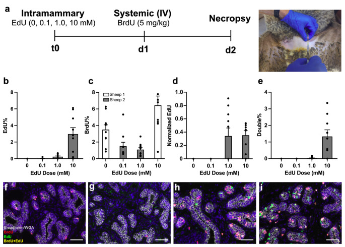
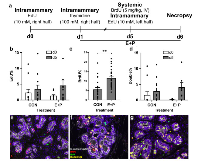
胸苷类似物(如乙炔基脱氧尿苷(EdU)或溴脱氧尿苷(BrdU))可用于标记乳腺上皮细胞(MEC)的有丝分裂,并对其增殖进行量化。然而,在大型动物体内标记细胞需要大量的化学物质,成本高且危险。我们开发了一种策略,将 EdU 注入母羊乳腺,直接标记有丝分裂的 MEC。首先,将四种不同浓度的 EdU(0、0.1、1.0 或 10 mM)中的一种注入空怀母羊(n = 2)的乳房内,24 小时后将其与 BrdU IV(5 mg/kg)进行比较。组织通过免疫荧光组织化学分析来检测 EdU、BrdU 和总 MEC。在测试的 EdU 剂量中,10 mM EdU 的标记指数最高,而一部分 MEC 同时被 EdU 和 BrdU 标记。接下来,我们试图确定乳腺内标记是否能检测暴露于外源性雌激素和孕酮(E+P)后有丝分裂的诱导。我们首先在t 0时将EdU(10 mM)注入母羊(n = 6)的右半乳房,然后在24小时后注入胸腺嘧啶(100 mM)以防止进一步标记。然后给 3 只母羊注射 E + P 5 d,n = 3 只母羊作为对照组。第 5 天,将 EdU 注入所有乳腺的左半乳房,同时静脉注射 BrdU(5 毫克/千克)。第 5 天,在乳房左半部,对照组腺体的最终 EdU 标记指数为 3.4%,而暴露于 E + P 的腺体的标记指数为 4.6%(p = 0.05)。CON腺体相应的BrdU标记指数为5.6%,而E+P后为12%(p = 0.05)。
Intramammary Labeling of Epithelial Cell Division.
Thymidine analogs such as ethynyl deoxyuridine (EdU) or bromodeoxyuridine (BrdU) can be used to label mitosis of mammary epithelial cells (MEC) and to quantify their proliferation. However, labeling cells in larger animals requires considerable amounts of chemical that can be costly and hazardous. We developed a strategy to infuse EdU into the mammary glands of ewes to directly label mitotic MEC. First, each udder half of nulliparous ewes (n = 2) received an intramammary infusion of one of four different concentrations of EdU (0, 0.1, 1.0 or 10 mM) which was compared to BrdU IV (5 mg/kg) 24 h later. Tissues were analyzed by immunofluorescent histochemistry to detect EdU, BrdU, and total MEC. Of the EdU doses tested, 10 mM EdU yielded the greatest labeling index, while a proportion of MEC were labeled by both EdU and BrdU. We next sought to establish whether intramammary labeling could detect the induction of mitosis after exposure to exogenous estrogen and progesterone (E + P). We first infused EdU (10 mM) into the right udder half of ewes (n = 6) at t 0, followed by thymidine (100 mM) 24 h later to prevent further labeling. Three ewes were then administered E + P for 5 d, while n = 3 ewes served as controls. On d 5, EdU was infused into the left udder half of all mammary glands alongside BrdU IV (5 mg/kg). By the time of necropsy 24 h later an average MEC labeling index of 2.9% resulted from EdU delivered at t 0. In the left half of the udder on d 5, CON glands had a final EdU labeling index of 3.4% while glands exposed to E + P had a labeling index of 4.6% (p = 0.05). The corresponding degree of labeling with BrdU was 5.6% in CON glands, and 12% following E + P (p < 0.001). Our findings reveal that intramammary labeling is an efficient and cost-effective method for single- and dual-labeling of cell division in the mammary glands.
期刊介绍:
Journal of Mammary Gland Biology and Neoplasia is the leading Journal in the field of mammary gland biology that provides researchers within and outside the field of mammary gland biology with an integrated source of information pertaining to the development, function, and pathology of the mammary gland and its function.
Commencing in 2015, the Journal will begin receiving and publishing a combination of reviews and original, peer-reviewed research. The Journal covers all topics related to the field of mammary gland biology, including mammary development, breast cancer biology, lactation, and milk composition and quality. The environmental, endocrine, nutritional, and molecular factors regulating these processes is covered, including from a comparative biology perspective.
 求助内容:
求助内容: 应助结果提醒方式:


