{"title":"研究阿尔茨海默氏症连续性痴呆症中 GAP-43 浓度与弥散张量成像指数之间的关联。","authors":"Armin Ariaei, Atousa Ghorbani, Elham Habibzadeh, Nazanin Moghaddam, Negar Chegeni Nezhad, Amirabbas Abdoli, Samira Mazinanian, Mohammad Sadeghi, Mahsa Mayeli","doi":"10.1186/s12883-024-03904-9","DOIUrl":null,"url":null,"abstract":"<p><strong>Background: </strong>Synaptic degeneration, axonal injury, and white matter disintegration are among the pathological events in Alzheimer's disease (AD), for which growth-associated protein 43 (GAP-43) and diffusion tensor imaging (DTI) could be an indicator. In this study, the cerebrospinal fluid (CSF) GAP-43 clinical trajectories and their association with progression and AD hallmarks with white matter microstructural changes were evaluated.</p><p><strong>Methods: </strong>A total number of 133 participants were enrolled in GAP-43 and DTI values were compared between groups, both cross-sectionally and longitudinally with two and four-year follow-ups. Subsequently, the correlation between GAP-43 levels in the CSF and DTI values was investigated using Spearman's correlation.</p><p><strong>Results: </strong>The CSF level of GAP-43 is negatively correlated with the mean diffusivity measures in Fornix (Cres)/Stria terminals in early and late MCI (r<sub>s</sub>=-0.478 p = 0.021 and r<sub>s</sub>=-0.425 p = 0.038). Additionally, the CSF level of GAP-43 is negatively correlated with fractional anisotropy in the cingulum in late MCI (r<sub>s</sub>=-0.437 p = 0.033). Moreover, the axial diffusivity in superior corona radiate (r<sub>s</sub>=-0.562 p = 0.005 and r<sub>s</sub>=-0.484 p = 0.036) and radial diffusivity in superior fronto-occipital fasciculus was negatively correlated with GAP-43 level in the early and mid-MCI participants (r<sub>s</sub>=-0.520 p = 0.011 and r<sub>s</sub>=-0.498 p = 0.030).</p><p><strong>Conclusions: </strong>Presynaptic marker GAP-43 in combination with DTI can be used as a novel biomarker to identify microstructural synaptic degeneration in the early MCI. In addition, it can be used as a biomarker for tracking the progression of AD and monitoring treatment efficacy.</p>","PeriodicalId":9170,"journal":{"name":"BMC Neurology","volume":"24 1","pages":"397"},"PeriodicalIF":2.2000,"publicationDate":"2024-10-17","publicationTypes":"Journal Article","fieldsOfStudy":null,"isOpenAccess":false,"openAccessPdf":"https://www.ncbi.nlm.nih.gov/pmc/articles/PMC11484424/pdf/","citationCount":"0","resultStr":"{\"title\":\"Investigating the association between the GAP-43 concentration with diffusion tensor imaging indices in Alzheimer's dementia continuum.\",\"authors\":\"Armin Ariaei, Atousa Ghorbani, Elham Habibzadeh, Nazanin Moghaddam, Negar Chegeni Nezhad, Amirabbas Abdoli, Samira Mazinanian, Mohammad Sadeghi, Mahsa Mayeli\",\"doi\":\"10.1186/s12883-024-03904-9\",\"DOIUrl\":null,\"url\":null,\"abstract\":\"<p><strong>Background: </strong>Synaptic degeneration, axonal injury, and white matter disintegration are among the pathological events in Alzheimer's disease (AD), for which growth-associated protein 43 (GAP-43) and diffusion tensor imaging (DTI) could be an indicator. In this study, the cerebrospinal fluid (CSF) GAP-43 clinical trajectories and their association with progression and AD hallmarks with white matter microstructural changes were evaluated.</p><p><strong>Methods: </strong>A total number of 133 participants were enrolled in GAP-43 and DTI values were compared between groups, both cross-sectionally and longitudinally with two and four-year follow-ups. Subsequently, the correlation between GAP-43 levels in the CSF and DTI values was investigated using Spearman's correlation.</p><p><strong>Results: </strong>The CSF level of GAP-43 is negatively correlated with the mean diffusivity measures in Fornix (Cres)/Stria terminals in early and late MCI (r<sub>s</sub>=-0.478 p = 0.021 and r<sub>s</sub>=-0.425 p = 0.038). Additionally, the CSF level of GAP-43 is negatively correlated with fractional anisotropy in the cingulum in late MCI (r<sub>s</sub>=-0.437 p = 0.033). Moreover, the axial diffusivity in superior corona radiate (r<sub>s</sub>=-0.562 p = 0.005 and r<sub>s</sub>=-0.484 p = 0.036) and radial diffusivity in superior fronto-occipital fasciculus was negatively correlated with GAP-43 level in the early and mid-MCI participants (r<sub>s</sub>=-0.520 p = 0.011 and r<sub>s</sub>=-0.498 p = 0.030).</p><p><strong>Conclusions: </strong>Presynaptic marker GAP-43 in combination with DTI can be used as a novel biomarker to identify microstructural synaptic degeneration in the early MCI. In addition, it can be used as a biomarker for tracking the progression of AD and monitoring treatment efficacy.</p>\",\"PeriodicalId\":9170,\"journal\":{\"name\":\"BMC Neurology\",\"volume\":\"24 1\",\"pages\":\"397\"},\"PeriodicalIF\":2.2000,\"publicationDate\":\"2024-10-17\",\"publicationTypes\":\"Journal Article\",\"fieldsOfStudy\":null,\"isOpenAccess\":false,\"openAccessPdf\":\"https://www.ncbi.nlm.nih.gov/pmc/articles/PMC11484424/pdf/\",\"citationCount\":\"0\",\"resultStr\":null,\"platform\":\"Semanticscholar\",\"paperid\":null,\"PeriodicalName\":\"BMC Neurology\",\"FirstCategoryId\":\"3\",\"ListUrlMain\":\"https://doi.org/10.1186/s12883-024-03904-9\",\"RegionNum\":3,\"RegionCategory\":\"医学\",\"ArticlePicture\":[],\"TitleCN\":null,\"AbstractTextCN\":null,\"PMCID\":null,\"EPubDate\":\"\",\"PubModel\":\"\",\"JCR\":\"Q3\",\"JCRName\":\"CLINICAL NEUROLOGY\",\"Score\":null,\"Total\":0}","platform":"Semanticscholar","paperid":null,"PeriodicalName":"BMC Neurology","FirstCategoryId":"3","ListUrlMain":"https://doi.org/10.1186/s12883-024-03904-9","RegionNum":3,"RegionCategory":"医学","ArticlePicture":[],"TitleCN":null,"AbstractTextCN":null,"PMCID":null,"EPubDate":"","PubModel":"","JCR":"Q3","JCRName":"CLINICAL NEUROLOGY","Score":null,"Total":0}
引用次数: 0
摘要
背景:突触变性、轴突损伤和白质破坏是阿尔茨海默病(AD)的病理特征之一,而生长相关蛋白 43(GAP-43)和弥散张量成像(DTI)可作为其指标。本研究评估了脑脊液(CSF)GAP-43的临床轨迹及其与进展和AD特征与白质微结构变化的关系:方法: 共有 133 名参与者参加了 GAP-43 研究,并在两年和四年的随访中横向和纵向比较了各组之间的 DTI 值。随后,利用斯皮尔曼相关性研究了脑脊液中 GAP-43 水平与 DTI 值之间的相关性:结果:GAP-43的CSF水平与早期和晚期MCI患者Fornix (Cres)/Stria末端的平均弥散度呈负相关(rs=-0.478 p = 0.021和rs=-0.425 p = 0.038)。此外,在晚期 MCI 中,GAP-43 的 CSF 水平与脑室各向异性分数呈负相关(rs=-0.437 p = 0.033)。此外,在早期和中期MCI患者中,放射状上冠的轴向扩散率(rs=-0.562 p = 0.005和rs=-0.484 p = 0.036)和枕前上筋膜的径向扩散率与GAP-43水平呈负相关(rs=-0.520 p = 0.011和rs=-0.498 p = 0.030):结论:突触前标记物GAP-43与DTI相结合可作为一种新型生物标记物,用于识别早期MCI患者的微结构突触退化。结论:突触前标记物 GAP-43 与 DTI 结合可作为一种新型生物标记物,用于识别早期 MCI 的微结构突触退化,此外,它还可作为一种生物标记物,用于追踪 AD 的进展和监测治疗效果。
Investigating the association between the GAP-43 concentration with diffusion tensor imaging indices in Alzheimer's dementia continuum.
Background: Synaptic degeneration, axonal injury, and white matter disintegration are among the pathological events in Alzheimer's disease (AD), for which growth-associated protein 43 (GAP-43) and diffusion tensor imaging (DTI) could be an indicator. In this study, the cerebrospinal fluid (CSF) GAP-43 clinical trajectories and their association with progression and AD hallmarks with white matter microstructural changes were evaluated.
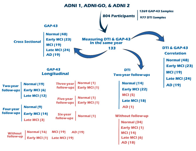
Methods: A total number of 133 participants were enrolled in GAP-43 and DTI values were compared between groups, both cross-sectionally and longitudinally with two and four-year follow-ups. Subsequently, the correlation between GAP-43 levels in the CSF and DTI values was investigated using Spearman's correlation.
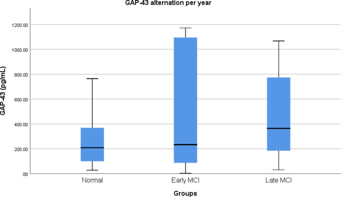
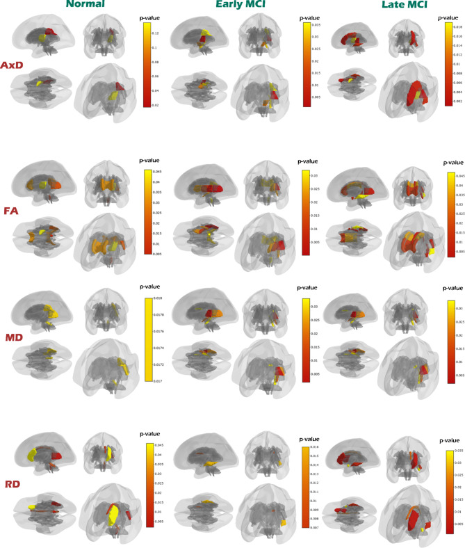
Results: The CSF level of GAP-43 is negatively correlated with the mean diffusivity measures in Fornix (Cres)/Stria terminals in early and late MCI (rs=-0.478 p = 0.021 and rs=-0.425 p = 0.038). Additionally, the CSF level of GAP-43 is negatively correlated with fractional anisotropy in the cingulum in late MCI (rs=-0.437 p = 0.033). Moreover, the axial diffusivity in superior corona radiate (rs=-0.562 p = 0.005 and rs=-0.484 p = 0.036) and radial diffusivity in superior fronto-occipital fasciculus was negatively correlated with GAP-43 level in the early and mid-MCI participants (rs=-0.520 p = 0.011 and rs=-0.498 p = 0.030).
Conclusions: Presynaptic marker GAP-43 in combination with DTI can be used as a novel biomarker to identify microstructural synaptic degeneration in the early MCI. In addition, it can be used as a biomarker for tracking the progression of AD and monitoring treatment efficacy.
期刊介绍:
BMC Neurology is an open access, peer-reviewed journal that considers articles on all aspects of the prevention, diagnosis and management of neurological disorders, as well as related molecular genetics, pathophysiology, and epidemiology.

 求助内容:
求助内容: 应助结果提醒方式:


