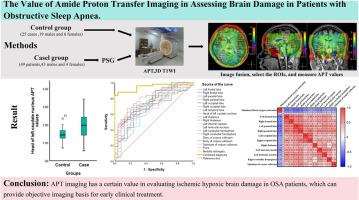
酰胺质子转移成像在评估阻塞性睡眠呼吸暂停患者脑损伤方面的价值

IF 3.8 2区 医学 Q1 CLINICAL NEUROLOGY
Ning Wang , Li Zhang , Jihua Zhang , Rongrong Zhang , Yan Zhao , Baogen Zhao , Shichao Wu , Ruifeng Zhang , Zeqiang Ji , Licun Lei , Wenyuan Wang , Yong Wang
{"title":"酰胺质子转移成像在评估阻塞性睡眠呼吸暂停患者脑损伤方面的价值","authors":"Ning Wang ,&nbsp;Li Zhang ,&nbsp;Jihua Zhang ,&nbsp;Rongrong Zhang ,&nbsp;Yan Zhao ,&nbsp;Baogen Zhao ,&nbsp;Shichao Wu ,&nbsp;Ruifeng Zhang ,&nbsp;Zeqiang Ji ,&nbsp;Licun Lei ,&nbsp;Wenyuan Wang ,&nbsp;Yong Wang","doi":"10.1016/j.sleep.2024.10.009","DOIUrl":null,"url":null,"abstract":"<div><h3>Objective</h3><div>To investigate the application value of amide proton transfer (APT) magnetic resonance imaging in evaluating brain damage in patients with obstructive sleep apnea (OSA).</div></div><div><h3>Materials and methods</h3><div>49 OSA patients and 25 healthy individuals matched for age and sex were included as case and control groups. All study participants underwent conventional 3D T1WI and APT imaging of the head. The APT values were measured in each of the brain regions of interest (ROI). To compare the differences in APT values of each brain region between the case and control groups, to compare the differences in the efficacy of APT values of each brain region in diagnosing OSA, and to investigate the correlation between APT values of each brain region and Apnea Hypopnea Index (AHI) and minimum blood oxygen saturation.</div></div><div><h3>Results</h3><div>Compared to the control group, patients with OSA had elevated APT values in several brain regions (P &lt; 0.05), and the diagnostic efficacy of the combined diagnosis of OSA by multiple brain regions is better than that by each brain region alone.Left frontal APT values were positively correlated with AHI in the case group (r = 0.33, P = 0.020).In the case group, the APT values of bilateral frontal lobe, parietal lobe, occipital lobe, left temporal lobe, right thalamus, left internal capsule, left lenticular nucleus, right cerebellar hemisphere and splenium of corpus callosum were negatively correlated with the minimum blood oxygen saturation, and the APT values for each of the above ROIs were positively correlated.</div></div><div><h3>Conclusion</h3><div>APT imaging has a certain value in evaluating brain damage in OSA patients, which may provide a new objective imaging basis for the clinical diagnosis of OSA.</div></div>","PeriodicalId":21874,"journal":{"name":"Sleep medicine","volume":"124 ","pages":"Pages 434-442"},"PeriodicalIF":3.8000,"publicationDate":"2024-10-10","publicationTypes":"Journal Article","fieldsOfStudy":null,"isOpenAccess":false,"openAccessPdf":"","citationCount":"0","resultStr":"{\"title\":\"The value of amide proton transfer imaging in assessing brain damage in patients with obstructive sleep apnea\",\"authors\":\"Ning Wang ,&nbsp;Li Zhang ,&nbsp;Jihua Zhang ,&nbsp;Rongrong Zhang ,&nbsp;Yan Zhao ,&nbsp;Baogen Zhao ,&nbsp;Shichao Wu ,&nbsp;Ruifeng Zhang ,&nbsp;Zeqiang Ji ,&nbsp;Licun Lei ,&nbsp;Wenyuan Wang ,&nbsp;Yong Wang\",\"doi\":\"10.1016/j.sleep.2024.10.009\",\"DOIUrl\":null,\"url\":null,\"abstract\":\"<div><h3>Objective</h3><div>To investigate the application value of amide proton transfer (APT) magnetic resonance imaging in evaluating brain damage in patients with obstructive sleep apnea (OSA).</div></div><div><h3>Materials and methods</h3><div>49 OSA patients and 25 healthy individuals matched for age and sex were included as case and control groups. All study participants underwent conventional 3D T1WI and APT imaging of the head. The APT values were measured in each of the brain regions of interest (ROI). To compare the differences in APT values of each brain region between the case and control groups, to compare the differences in the efficacy of APT values of each brain region in diagnosing OSA, and to investigate the correlation between APT values of each brain region and Apnea Hypopnea Index (AHI) and minimum blood oxygen saturation.</div></div><div><h3>Results</h3><div>Compared to the control group, patients with OSA had elevated APT values in several brain regions (P &lt; 0.05), and the diagnostic efficacy of the combined diagnosis of OSA by multiple brain regions is better than that by each brain region alone.Left frontal APT values were positively correlated with AHI in the case group (r = 0.33, P = 0.020).In the case group, the APT values of bilateral frontal lobe, parietal lobe, occipital lobe, left temporal lobe, right thalamus, left internal capsule, left lenticular nucleus, right cerebellar hemisphere and splenium of corpus callosum were negatively correlated with the minimum blood oxygen saturation, and the APT values for each of the above ROIs were positively correlated.</div></div><div><h3>Conclusion</h3><div>APT imaging has a certain value in evaluating brain damage in OSA patients, which may provide a new objective imaging basis for the clinical diagnosis of OSA.</div></div>\",\"PeriodicalId\":21874,\"journal\":{\"name\":\"Sleep medicine\",\"volume\":\"124 \",\"pages\":\"Pages 434-442\"},\"PeriodicalIF\":3.8000,\"publicationDate\":\"2024-10-10\",\"publicationTypes\":\"Journal Article\",\"fieldsOfStudy\":null,\"isOpenAccess\":false,\"openAccessPdf\":\"\",\"citationCount\":\"0\",\"resultStr\":null,\"platform\":\"Semanticscholar\",\"paperid\":null,\"PeriodicalName\":\"Sleep medicine\",\"FirstCategoryId\":\"3\",\"ListUrlMain\":\"https://www.sciencedirect.com/science/article/pii/S1389945724004751\",\"RegionNum\":2,\"RegionCategory\":\"医学\",\"ArticlePicture\":[],\"TitleCN\":null,\"AbstractTextCN\":null,\"PMCID\":null,\"EPubDate\":\"\",\"PubModel\":\"\",\"JCR\":\"Q1\",\"JCRName\":\"CLINICAL NEUROLOGY\",\"Score\":null,\"Total\":0}","platform":"Semanticscholar","paperid":null,"PeriodicalName":"Sleep medicine","FirstCategoryId":"3","ListUrlMain":"https://www.sciencedirect.com/science/article/pii/S1389945724004751","RegionNum":2,"RegionCategory":"医学","ArticlePicture":[],"TitleCN":null,"AbstractTextCN":null,"PMCID":null,"EPubDate":"","PubModel":"","JCR":"Q1","JCRName":"CLINICAL NEUROLOGY","Score":null,"Total":0}
引用次数: 0

摘要

目的 探讨酰胺质子转移(APT)磁共振成像在评估阻塞性睡眠呼吸暂停(OSA)患者脑损伤中的应用价值。所有研究参与者均接受了头部常规三维 T1WI 和 APT 成像检查。每个大脑感兴趣区(ROI)都测量了 APT 值。比较病例组和对照组各脑区 APT 值的差异,比较各脑区 APT 值在诊断 OSA 方面的疗效差异,并研究各脑区 APT 值与呼吸暂停低通气指数(AHI)和最低血氧饱和度之间的相关性。结果与对照组相比,OSA患者多个脑区的APT值升高(P <0.05),多脑区联合诊断OSA的疗效优于各脑区单独诊断。病例组的左额叶 APT 值与 AHI 呈正相关(r = 0.33,P = 0.020)。在病例组中,双侧额叶、顶叶、枕叶、左颞叶、右丘脑、左内囊、左扁桃体核、右小脑半球和胼胝体脾的 APT 值与最低血氧饱和度呈负相关,上述各 ROI 的 APT 值呈正相关。结论 APT成像在评价OSA患者脑损伤方面具有一定价值,可为临床诊断OSA提供新的客观成像依据。
本文章由计算机程序翻译,如有差异,请以英文原文为准。

The value of amide proton transfer imaging in assessing brain damage in patients with obstructive sleep apnea

The value of amide proton transfer imaging in assessing brain damage in patients with obstructive sleep apnea

Objective

To investigate the application value of amide proton transfer (APT) magnetic resonance imaging in evaluating brain damage in patients with obstructive sleep apnea (OSA).

Materials and methods

49 OSA patients and 25 healthy individuals matched for age and sex were included as case and control groups. All study participants underwent conventional 3D T1WI and APT imaging of the head. The APT values were measured in each of the brain regions of interest (ROI). To compare the differences in APT values of each brain region between the case and control groups, to compare the differences in the efficacy of APT values of each brain region in diagnosing OSA, and to investigate the correlation between APT values of each brain region and Apnea Hypopnea Index (AHI) and minimum blood oxygen saturation.

Results

Compared to the control group, patients with OSA had elevated APT values in several brain regions (P < 0.05), and the diagnostic efficacy of the combined diagnosis of OSA by multiple brain regions is better than that by each brain region alone.Left frontal APT values were positively correlated with AHI in the case group (r = 0.33, P = 0.020).In the case group, the APT values of bilateral frontal lobe, parietal lobe, occipital lobe, left temporal lobe, right thalamus, left internal capsule, left lenticular nucleus, right cerebellar hemisphere and splenium of corpus callosum were negatively correlated with the minimum blood oxygen saturation, and the APT values for each of the above ROIs were positively correlated.

Conclusion

APT imaging has a certain value in evaluating brain damage in OSA patients, which may provide a new objective imaging basis for the clinical diagnosis of OSA.
求助全文
通过发布文献求助,成功后即可免费获取论文全文。 去求助
来源期刊
Sleep medicine
Sleep medicine 医学-临床神经学
CiteScore
8.40
自引率
6.20%
发文量
1060
审稿时长
49 days
期刊介绍: Sleep Medicine aims to be a journal no one involved in clinical sleep medicine can do without. A journal primarily focussing on the human aspects of sleep, integrating the various disciplines that are involved in sleep medicine: neurology, clinical neurophysiology, internal medicine (particularly pulmonology and cardiology), psychology, psychiatry, sleep technology, pediatrics, neurosurgery, otorhinolaryngology, and dentistry. The journal publishes the following types of articles: Reviews (also intended as a way to bridge the gap between basic sleep research and clinical relevance); Original Research Articles; Full-length articles; Brief communications; Controversies; Case reports; Letters to the Editor; Journal search and commentaries; Book reviews; Meeting announcements; Listing of relevant organisations plus web sites.
×
引用
GB/T 7714-2015
复制
MLA
复制
APA
复制
导出至
BibTeX EndNote RefMan NoteFirst NoteExpress
×
提示
您的信息不完整,为了账户安全,请先补充。
现在去补充
×
提示
您因"违规操作"
具体请查看互助需知
我知道了
×
提示
确定
请完成安全验证×
copy
已复制链接
快去分享给好友吧!
我知道了
右上角分享
点击右上角分享
0
联系我们:info@booksci.cn Book学术提供免费学术资源搜索服务,方便国内外学者检索中英文文献。致力于提供最便捷和优质的服务体验。 Copyright © 2023 布克学术 All rights reserved.
京ICP备2023020795号-1
ghs 京公网安备 11010802042870号
Book学术文献互助
Book学术文献互助群
群 号:481959085
Book学术官方微信