Christopher Thomas, Tabea Lilian Marx, Sarah Mae Penir, Melina Schuh
{"title":"体外成像揭示排卵的时空控制","authors":"Christopher Thomas, Tabea Lilian Marx, Sarah Mae Penir, Melina Schuh","doi":"10.1038/s41556-024-01524-6","DOIUrl":null,"url":null,"abstract":"During ovulation, an egg is released from an ovarian follicle, ready for fertilization. Ovulation occurs inside the body, impeding direct studies of its progression. Therefore, the exact mechanisms that control ovulation have remained unclear. Here we devised live imaging methods to study the entire process of ovulation in isolated mouse ovarian follicles. We show that ovulation proceeds through three distinct phases, follicle expansion (I), contraction (II) and rupture (III), culminating in the release of the egg. Follicle expansion is driven by hyaluronic acid secretion and an osmotic gradient-directed fluid influx into the follicle. Then, smooth muscle cells in the outer follicle drive follicle contraction. Follicle rupture begins with stigma formation, followed by the exit of follicular fluid and cumulus cells and the rapid release of the egg. These results establish a mechanistic framework for ovulation, a process of fundamental importance for reproduction. Thomas, Marx et al. devise a live imaging approach to spatiotemporally dissect mouse ovulation ex vivo.","PeriodicalId":18977,"journal":{"name":"Nature Cell Biology","volume":"26 11","pages":"1997-2008"},"PeriodicalIF":17.3000,"publicationDate":"2024-10-16","publicationTypes":"Journal Article","fieldsOfStudy":null,"isOpenAccess":false,"openAccessPdf":"https://www.nature.com/articles/s41556-024-01524-6.pdf","citationCount":"0","resultStr":"{\"title\":\"Ex vivo imaging reveals the spatiotemporal control of ovulation\",\"authors\":\"Christopher Thomas, Tabea Lilian Marx, Sarah Mae Penir, Melina Schuh\",\"doi\":\"10.1038/s41556-024-01524-6\",\"DOIUrl\":null,\"url\":null,\"abstract\":\"During ovulation, an egg is released from an ovarian follicle, ready for fertilization. Ovulation occurs inside the body, impeding direct studies of its progression. Therefore, the exact mechanisms that control ovulation have remained unclear. Here we devised live imaging methods to study the entire process of ovulation in isolated mouse ovarian follicles. We show that ovulation proceeds through three distinct phases, follicle expansion (I), contraction (II) and rupture (III), culminating in the release of the egg. Follicle expansion is driven by hyaluronic acid secretion and an osmotic gradient-directed fluid influx into the follicle. Then, smooth muscle cells in the outer follicle drive follicle contraction. Follicle rupture begins with stigma formation, followed by the exit of follicular fluid and cumulus cells and the rapid release of the egg. These results establish a mechanistic framework for ovulation, a process of fundamental importance for reproduction. Thomas, Marx et al. devise a live imaging approach to spatiotemporally dissect mouse ovulation ex vivo.\",\"PeriodicalId\":18977,\"journal\":{\"name\":\"Nature Cell Biology\",\"volume\":\"26 11\",\"pages\":\"1997-2008\"},\"PeriodicalIF\":17.3000,\"publicationDate\":\"2024-10-16\",\"publicationTypes\":\"Journal Article\",\"fieldsOfStudy\":null,\"isOpenAccess\":false,\"openAccessPdf\":\"https://www.nature.com/articles/s41556-024-01524-6.pdf\",\"citationCount\":\"0\",\"resultStr\":null,\"platform\":\"Semanticscholar\",\"paperid\":null,\"PeriodicalName\":\"Nature Cell Biology\",\"FirstCategoryId\":\"99\",\"ListUrlMain\":\"https://www.nature.com/articles/s41556-024-01524-6\",\"RegionNum\":1,\"RegionCategory\":\"生物学\",\"ArticlePicture\":[],\"TitleCN\":null,\"AbstractTextCN\":null,\"PMCID\":null,\"EPubDate\":\"\",\"PubModel\":\"\",\"JCR\":\"Q1\",\"JCRName\":\"CELL BIOLOGY\",\"Score\":null,\"Total\":0}","platform":"Semanticscholar","paperid":null,"PeriodicalName":"Nature Cell Biology","FirstCategoryId":"99","ListUrlMain":"https://www.nature.com/articles/s41556-024-01524-6","RegionNum":1,"RegionCategory":"生物学","ArticlePicture":[],"TitleCN":null,"AbstractTextCN":null,"PMCID":null,"EPubDate":"","PubModel":"","JCR":"Q1","JCRName":"CELL BIOLOGY","Score":null,"Total":0}
Ex vivo imaging reveals the spatiotemporal control of ovulation
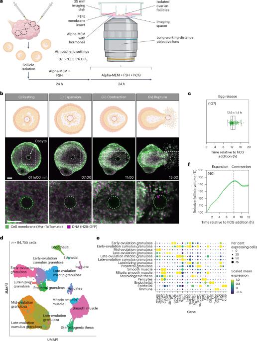
During ovulation, an egg is released from an ovarian follicle, ready for fertilization. Ovulation occurs inside the body, impeding direct studies of its progression. Therefore, the exact mechanisms that control ovulation have remained unclear. Here we devised live imaging methods to study the entire process of ovulation in isolated mouse ovarian follicles. We show that ovulation proceeds through three distinct phases, follicle expansion (I), contraction (II) and rupture (III), culminating in the release of the egg. Follicle expansion is driven by hyaluronic acid secretion and an osmotic gradient-directed fluid influx into the follicle. Then, smooth muscle cells in the outer follicle drive follicle contraction. Follicle rupture begins with stigma formation, followed by the exit of follicular fluid and cumulus cells and the rapid release of the egg. These results establish a mechanistic framework for ovulation, a process of fundamental importance for reproduction. Thomas, Marx et al. devise a live imaging approach to spatiotemporally dissect mouse ovulation ex vivo.
期刊介绍:
Nature Cell Biology, a prestigious journal, upholds a commitment to publishing papers of the highest quality across all areas of cell biology, with a particular focus on elucidating mechanisms underlying fundamental cell biological processes. The journal's broad scope encompasses various areas of interest, including but not limited to:
-Autophagy
-Cancer biology
-Cell adhesion and migration
-Cell cycle and growth
-Cell death
-Chromatin and epigenetics
-Cytoskeletal dynamics
-Developmental biology
-DNA replication and repair
-Mechanisms of human disease
-Mechanobiology
-Membrane traffic and dynamics
-Metabolism
-Nuclear organization and dynamics
-Organelle biology
-Proteolysis and quality control
-RNA biology
-Signal transduction
-Stem cell biology