Erli Cai, Yage Chen, Jing Zhang, Haozheng Li, Yiran Li, Shuai Yan, Zhiyong He, Quan Yuan and Ping Wang
{"title":"利用附有非天然氨基酸的小型拉曼报告器对活细胞中的特定蛋白质进行成像","authors":"Erli Cai, Yage Chen, Jing Zhang, Haozheng Li, Yiran Li, Shuai Yan, Zhiyong He, Quan Yuan and Ping Wang","doi":"10.1039/D4AN00758A","DOIUrl":null,"url":null,"abstract":"<p >Fluorescence labeling <em>via</em> fluorescent proteins (FPs) or immunofluorescence has been routinely applied for microscopic imaging of specific proteins. However, due to these over-weight and oversized labels (<em>e.g.</em> GFP, 238 aa, 27 kDa, ∼4 nm in size), the potential physiological malfunctions of the target proteins are largely underestimated in living cells. Herein, for living cells, we report a small and minimally-invasive Raman reporter (about 2 aa and <1 kDa), which can be site-specifically introduced into proteins by genetic codon expansion. After a single unnatural amino acid (UAA) is precisely incorporated into the target protein, the strained alkyne can rapidly undergo copper-free Diels–Alder cycloaddition reactions with the tetrazine-functionalized Raman reporter, which features a fine vibrational spectrum in contrast to fluorescence. In our experimental results, the UAA-based Raman tag was successfully incorporated into vimentin, histone 3.3 and huntingtin (Htt74Q) proteins in living HeLa cells and further utilized for stimulated Raman imaging. The site-specific bioorthogonal fusion of small Raman tags with intracellular proteins will pave the way for minimally-invasive protein labeling and multi-color imaging in living cells.</p>","PeriodicalId":63,"journal":{"name":"Analyst","volume":" 22","pages":" 5476-5481"},"PeriodicalIF":3.6000,"publicationDate":"2024-10-14","publicationTypes":"Journal Article","fieldsOfStudy":null,"isOpenAccess":false,"openAccessPdf":"","citationCount":"0","resultStr":"{\"title\":\"Imaging specific proteins in living cells with small unnatural amino acid attached Raman reporters†\",\"authors\":\"Erli Cai, Yage Chen, Jing Zhang, Haozheng Li, Yiran Li, Shuai Yan, Zhiyong He, Quan Yuan and Ping Wang\",\"doi\":\"10.1039/D4AN00758A\",\"DOIUrl\":null,\"url\":null,\"abstract\":\"<p >Fluorescence labeling <em>via</em> fluorescent proteins (FPs) or immunofluorescence has been routinely applied for microscopic imaging of specific proteins. However, due to these over-weight and oversized labels (<em>e.g.</em> GFP, 238 aa, 27 kDa, ∼4 nm in size), the potential physiological malfunctions of the target proteins are largely underestimated in living cells. Herein, for living cells, we report a small and minimally-invasive Raman reporter (about 2 aa and <1 kDa), which can be site-specifically introduced into proteins by genetic codon expansion. After a single unnatural amino acid (UAA) is precisely incorporated into the target protein, the strained alkyne can rapidly undergo copper-free Diels–Alder cycloaddition reactions with the tetrazine-functionalized Raman reporter, which features a fine vibrational spectrum in contrast to fluorescence. In our experimental results, the UAA-based Raman tag was successfully incorporated into vimentin, histone 3.3 and huntingtin (Htt74Q) proteins in living HeLa cells and further utilized for stimulated Raman imaging. The site-specific bioorthogonal fusion of small Raman tags with intracellular proteins will pave the way for minimally-invasive protein labeling and multi-color imaging in living cells.</p>\",\"PeriodicalId\":63,\"journal\":{\"name\":\"Analyst\",\"volume\":\" 22\",\"pages\":\" 5476-5481\"},\"PeriodicalIF\":3.6000,\"publicationDate\":\"2024-10-14\",\"publicationTypes\":\"Journal Article\",\"fieldsOfStudy\":null,\"isOpenAccess\":false,\"openAccessPdf\":\"\",\"citationCount\":\"0\",\"resultStr\":null,\"platform\":\"Semanticscholar\",\"paperid\":null,\"PeriodicalName\":\"Analyst\",\"FirstCategoryId\":\"92\",\"ListUrlMain\":\"https://pubs.rsc.org/en/content/articlelanding/2024/an/d4an00758a\",\"RegionNum\":3,\"RegionCategory\":\"化学\",\"ArticlePicture\":[],\"TitleCN\":null,\"AbstractTextCN\":null,\"PMCID\":null,\"EPubDate\":\"\",\"PubModel\":\"\",\"JCR\":\"Q2\",\"JCRName\":\"CHEMISTRY, ANALYTICAL\",\"Score\":null,\"Total\":0}","platform":"Semanticscholar","paperid":null,"PeriodicalName":"Analyst","FirstCategoryId":"92","ListUrlMain":"https://pubs.rsc.org/en/content/articlelanding/2024/an/d4an00758a","RegionNum":3,"RegionCategory":"化学","ArticlePicture":[],"TitleCN":null,"AbstractTextCN":null,"PMCID":null,"EPubDate":"","PubModel":"","JCR":"Q2","JCRName":"CHEMISTRY, ANALYTICAL","Score":null,"Total":0}
引用次数: 0
摘要
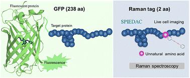
通过荧光蛋白(FPs)或免疫荧光进行荧光标记已被常规应用于特定蛋白质的显微成像。然而,由于这些标签重量过重、体积过大(如 GFP,238 aa,27 kDa,大小∼4 nm),目标蛋白质在活细胞中的潜在生理功能失常在很大程度上被低估了。在此,我们报告了一种针对活细胞的小型微创拉曼报告物(约 2 aa 和 1 kDa),该报告物可通过基因密码子扩增特异性地引入蛋白质中。将单个非天然氨基酸(UAA)精确地加入目标蛋白质后,受约束的炔烃可与四嗪功能化的拉曼报告物迅速发生无铜的 Diels-Alder 环加成反应,与荧光相比,拉曼报告物具有精细的振动光谱。根据我们的实验结果,基于 UAA 的拉曼标签被成功整合到活体 HeLa 细胞中的波形蛋白、组蛋白 3.3 和亨廷蛋白(Htt74Q)中,并进一步用于刺激拉曼成像。小型拉曼标签与细胞内蛋白质的特定位点生物正交融合将为活细胞中的微创蛋白质标记和多色成像铺平道路。
Imaging specific proteins in living cells with small unnatural amino acid attached Raman reporters†
Fluorescence labeling via fluorescent proteins (FPs) or immunofluorescence has been routinely applied for microscopic imaging of specific proteins. However, due to these over-weight and oversized labels (e.g. GFP, 238 aa, 27 kDa, ∼4 nm in size), the potential physiological malfunctions of the target proteins are largely underestimated in living cells. Herein, for living cells, we report a small and minimally-invasive Raman reporter (about 2 aa and <1 kDa), which can be site-specifically introduced into proteins by genetic codon expansion. After a single unnatural amino acid (UAA) is precisely incorporated into the target protein, the strained alkyne can rapidly undergo copper-free Diels–Alder cycloaddition reactions with the tetrazine-functionalized Raman reporter, which features a fine vibrational spectrum in contrast to fluorescence. In our experimental results, the UAA-based Raman tag was successfully incorporated into vimentin, histone 3.3 and huntingtin (Htt74Q) proteins in living HeLa cells and further utilized for stimulated Raman imaging. The site-specific bioorthogonal fusion of small Raman tags with intracellular proteins will pave the way for minimally-invasive protein labeling and multi-color imaging in living cells.

 求助内容:
求助内容: 应助结果提醒方式:


