Wenbin Liu , Kai Zhang , Yan Sun , Zhenyang Xiao , Hongkun Hu , Zixuan Xiong , Yihe Hu
{"title":"使用融合肽接枝壳聚糖涂层多孔钽支架多维治疗假体周围关节感染","authors":"Wenbin Liu , Kai Zhang , Yan Sun , Zhenyang Xiao , Hongkun Hu , Zixuan Xiong , Yihe Hu","doi":"10.1016/j.bioactmat.2024.09.040","DOIUrl":null,"url":null,"abstract":"<div><div>Bacterial infection and delayed osteointegration are two major challenges for orthopedic implants. Surface modification enables the implant have a time-sequenced biological function of effective antibacterial in the early stage and stable osteogenesis in the later stage, which is expected to achieve the purpose of preventing infection and prosthetic loosening after implant surgery. This study aims to construct a composite coating of carboxymethyl chitosan (CCS) grafted with an antibacterial (HHC36) and angiogenic (FP) fusion peptide (FP) on the surface of 3D-printed porous tantalum (Ta-CCS@FP) using alkaline treatment, electrostatic adsorption, and EDC/NHS reaction, to functionalize the surface coating while maintaining the original advantages of the material. The functionalized implants (Ta-CCS@FP) achieve sustained FP release in the initial stages, exhibiting potent antibacterial and anti-biofilm properties due to the synergistic action of the antimicrobial peptides (AMPs) HHC36 and CCS in disrupting bacterial membranes. Additionally, Ta-CCS@FP demonstrate robust osteogenic and angiogenic capabilities compared to Ta and Ta-CCS, attributed to QK and CCS. Notably, the conditioned medium intervention experiments of HUVECs and BMSCs showed that the implants had good angiogenic-osteogenic coupling properties. <em>In vivo</em> assays using infection bone defect models revealed that these bioactive implants effectively eradicated bacteria within 2 weeks and facilitated vascularized bone regeneration by 6 weeks. Thus, our study offers an integrated approach to address bacterial infection and enhance osseointegration for porous tantalum implants.</div></div>","PeriodicalId":8762,"journal":{"name":"Bioactive Materials","volume":"44 ","pages":"Pages 15-33"},"PeriodicalIF":18.0000,"publicationDate":"2024-10-11","publicationTypes":"Journal Article","fieldsOfStudy":null,"isOpenAccess":false,"openAccessPdf":"","citationCount":"0","resultStr":"{\"title\":\"Multidimensional treatment of periprosthetic joint infection using fusion peptide-grafted chitosan coated porous tantalum scaffold\",\"authors\":\"Wenbin Liu , Kai Zhang , Yan Sun , Zhenyang Xiao , Hongkun Hu , Zixuan Xiong , Yihe Hu\",\"doi\":\"10.1016/j.bioactmat.2024.09.040\",\"DOIUrl\":null,\"url\":null,\"abstract\":\"<div><div>Bacterial infection and delayed osteointegration are two major challenges for orthopedic implants. Surface modification enables the implant have a time-sequenced biological function of effective antibacterial in the early stage and stable osteogenesis in the later stage, which is expected to achieve the purpose of preventing infection and prosthetic loosening after implant surgery. This study aims to construct a composite coating of carboxymethyl chitosan (CCS) grafted with an antibacterial (HHC36) and angiogenic (FP) fusion peptide (FP) on the surface of 3D-printed porous tantalum (Ta-CCS@FP) using alkaline treatment, electrostatic adsorption, and EDC/NHS reaction, to functionalize the surface coating while maintaining the original advantages of the material. The functionalized implants (Ta-CCS@FP) achieve sustained FP release in the initial stages, exhibiting potent antibacterial and anti-biofilm properties due to the synergistic action of the antimicrobial peptides (AMPs) HHC36 and CCS in disrupting bacterial membranes. Additionally, Ta-CCS@FP demonstrate robust osteogenic and angiogenic capabilities compared to Ta and Ta-CCS, attributed to QK and CCS. Notably, the conditioned medium intervention experiments of HUVECs and BMSCs showed that the implants had good angiogenic-osteogenic coupling properties. <em>In vivo</em> assays using infection bone defect models revealed that these bioactive implants effectively eradicated bacteria within 2 weeks and facilitated vascularized bone regeneration by 6 weeks. Thus, our study offers an integrated approach to address bacterial infection and enhance osseointegration for porous tantalum implants.</div></div>\",\"PeriodicalId\":8762,\"journal\":{\"name\":\"Bioactive Materials\",\"volume\":\"44 \",\"pages\":\"Pages 15-33\"},\"PeriodicalIF\":18.0000,\"publicationDate\":\"2024-10-11\",\"publicationTypes\":\"Journal Article\",\"fieldsOfStudy\":null,\"isOpenAccess\":false,\"openAccessPdf\":\"\",\"citationCount\":\"0\",\"resultStr\":null,\"platform\":\"Semanticscholar\",\"paperid\":null,\"PeriodicalName\":\"Bioactive Materials\",\"FirstCategoryId\":\"5\",\"ListUrlMain\":\"https://www.sciencedirect.com/science/article/pii/S2452199X24004432\",\"RegionNum\":1,\"RegionCategory\":\"医学\",\"ArticlePicture\":[],\"TitleCN\":null,\"AbstractTextCN\":null,\"PMCID\":null,\"EPubDate\":\"\",\"PubModel\":\"\",\"JCR\":\"Q1\",\"JCRName\":\"ENGINEERING, BIOMEDICAL\",\"Score\":null,\"Total\":0}","platform":"Semanticscholar","paperid":null,"PeriodicalName":"Bioactive Materials","FirstCategoryId":"5","ListUrlMain":"https://www.sciencedirect.com/science/article/pii/S2452199X24004432","RegionNum":1,"RegionCategory":"医学","ArticlePicture":[],"TitleCN":null,"AbstractTextCN":null,"PMCID":null,"EPubDate":"","PubModel":"","JCR":"Q1","JCRName":"ENGINEERING, BIOMEDICAL","Score":null,"Total":0}
Multidimensional treatment of periprosthetic joint infection using fusion peptide-grafted chitosan coated porous tantalum scaffold
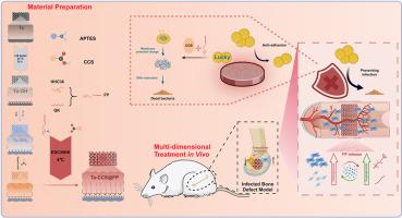
Bacterial infection and delayed osteointegration are two major challenges for orthopedic implants. Surface modification enables the implant have a time-sequenced biological function of effective antibacterial in the early stage and stable osteogenesis in the later stage, which is expected to achieve the purpose of preventing infection and prosthetic loosening after implant surgery. This study aims to construct a composite coating of carboxymethyl chitosan (CCS) grafted with an antibacterial (HHC36) and angiogenic (FP) fusion peptide (FP) on the surface of 3D-printed porous tantalum (Ta-CCS@FP) using alkaline treatment, electrostatic adsorption, and EDC/NHS reaction, to functionalize the surface coating while maintaining the original advantages of the material. The functionalized implants (Ta-CCS@FP) achieve sustained FP release in the initial stages, exhibiting potent antibacterial and anti-biofilm properties due to the synergistic action of the antimicrobial peptides (AMPs) HHC36 and CCS in disrupting bacterial membranes. Additionally, Ta-CCS@FP demonstrate robust osteogenic and angiogenic capabilities compared to Ta and Ta-CCS, attributed to QK and CCS. Notably, the conditioned medium intervention experiments of HUVECs and BMSCs showed that the implants had good angiogenic-osteogenic coupling properties. In vivo assays using infection bone defect models revealed that these bioactive implants effectively eradicated bacteria within 2 weeks and facilitated vascularized bone regeneration by 6 weeks. Thus, our study offers an integrated approach to address bacterial infection and enhance osseointegration for porous tantalum implants.
Bioactive MaterialsBiochemistry, Genetics and Molecular Biology-Biotechnology
CiteScore
28.00
自引率
6.30%
发文量
436
审稿时长
20 days
期刊介绍:
Bioactive Materials is a peer-reviewed research publication that focuses on advancements in bioactive materials. The journal accepts research papers, reviews, and rapid communications in the field of next-generation biomaterials that interact with cells, tissues, and organs in various living organisms.
The primary goal of Bioactive Materials is to promote the science and engineering of biomaterials that exhibit adaptiveness to the biological environment. These materials are specifically designed to stimulate or direct appropriate cell and tissue responses or regulate interactions with microorganisms.
The journal covers a wide range of bioactive materials, including those that are engineered or designed in terms of their physical form (e.g. particulate, fiber), topology (e.g. porosity, surface roughness), or dimensions (ranging from macro to nano-scales). Contributions are sought from the following categories of bioactive materials:
Bioactive metals and alloys
Bioactive inorganics: ceramics, glasses, and carbon-based materials
Bioactive polymers and gels
Bioactive materials derived from natural sources
Bioactive composites
These materials find applications in human and veterinary medicine, such as implants, tissue engineering scaffolds, cell/drug/gene carriers, as well as imaging and sensing devices.