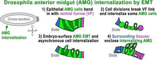
果蝇前中肠通过胚胎表面的集体上皮-间质转化和周围组织的包围而内化。

IF 2.5 3区 生物学 Q2 DEVELOPMENTAL BIOLOGY
Sandra Sabbagh, Hui Zhang, Tony J.C. Harris
{"title":"果蝇前中肠通过胚胎表面的集体上皮-间质转化和周围组织的包围而内化。","authors":"Sandra Sabbagh,&nbsp;Hui Zhang,&nbsp;Tony J.C. Harris","doi":"10.1016/j.ydbio.2024.10.002","DOIUrl":null,"url":null,"abstract":"<div><div>Internal organ development requires cell internalization, which can occur individually or collectively. The best characterized mode of collective internalization is epithelial invagination. Alternate modes involving collective mesenchymal behaviours at the embryo surface have been documented, but their prevalence is unclear. The Drosophila embryo has been a major model for the study of epithelial invaginations. However, internalization of the Drosophila anterior midgut primordium is incompletely understood. Here, we report that an epithelial-mesenchymal transition (EMT) occurs across the internalizing primordium when it is still at the embryo surface. At the earliest internalization stage, the primordium displays less junctional DE-cadherin than surrounding tissues but still exhibits coordinated epithelial structure as it invaginates with the ventral furrow. This initial invagination is transient, and its loss correlates with the activation of an associated mitotic domain. Activation of a subsequent mitotic domain across the broader primordium results in cell divisions with mixed orientations that deposit some cells within the embryo. However, cell division is non-essential for primordium internalization. Post-mitotically, the surface primordium displays hallmarks of EMT: loss of adherens junctions, loss of epithelial cell polarity, and gain of cell protrusions. Primordium cells extend over each other as they internalize asynchronously as individuals or small groups, and the primordium becomes enclosed by the reorganizations of surrounding epithelial tissues. We propose that collective EMT at the embryo surface promotes anterior midgut internalization through both inwardly-directed divisions and movements of its cells, and that the latter process is facilitated by surrounding tissue remodeling.</div></div>","PeriodicalId":11070,"journal":{"name":"Developmental biology","volume":"517 ","pages":"Pages 191-202"},"PeriodicalIF":2.5000,"publicationDate":"2024-10-09","publicationTypes":"Journal Article","fieldsOfStudy":null,"isOpenAccess":false,"openAccessPdf":"","citationCount":"0","resultStr":"{\"title\":\"Drosophila anterior midgut internalization via collective epithelial-mesenchymal transition at the embryo surface and enclosure by surrounding tissues\",\"authors\":\"Sandra Sabbagh,&nbsp;Hui Zhang,&nbsp;Tony J.C. Harris\",\"doi\":\"10.1016/j.ydbio.2024.10.002\",\"DOIUrl\":null,\"url\":null,\"abstract\":\"<div><div>Internal organ development requires cell internalization, which can occur individually or collectively. The best characterized mode of collective internalization is epithelial invagination. Alternate modes involving collective mesenchymal behaviours at the embryo surface have been documented, but their prevalence is unclear. The Drosophila embryo has been a major model for the study of epithelial invaginations. However, internalization of the Drosophila anterior midgut primordium is incompletely understood. Here, we report that an epithelial-mesenchymal transition (EMT) occurs across the internalizing primordium when it is still at the embryo surface. At the earliest internalization stage, the primordium displays less junctional DE-cadherin than surrounding tissues but still exhibits coordinated epithelial structure as it invaginates with the ventral furrow. This initial invagination is transient, and its loss correlates with the activation of an associated mitotic domain. Activation of a subsequent mitotic domain across the broader primordium results in cell divisions with mixed orientations that deposit some cells within the embryo. However, cell division is non-essential for primordium internalization. Post-mitotically, the surface primordium displays hallmarks of EMT: loss of adherens junctions, loss of epithelial cell polarity, and gain of cell protrusions. Primordium cells extend over each other as they internalize asynchronously as individuals or small groups, and the primordium becomes enclosed by the reorganizations of surrounding epithelial tissues. We propose that collective EMT at the embryo surface promotes anterior midgut internalization through both inwardly-directed divisions and movements of its cells, and that the latter process is facilitated by surrounding tissue remodeling.</div></div>\",\"PeriodicalId\":11070,\"journal\":{\"name\":\"Developmental biology\",\"volume\":\"517 \",\"pages\":\"Pages 191-202\"},\"PeriodicalIF\":2.5000,\"publicationDate\":\"2024-10-09\",\"publicationTypes\":\"Journal Article\",\"fieldsOfStudy\":null,\"isOpenAccess\":false,\"openAccessPdf\":\"\",\"citationCount\":\"0\",\"resultStr\":null,\"platform\":\"Semanticscholar\",\"paperid\":null,\"PeriodicalName\":\"Developmental biology\",\"FirstCategoryId\":\"99\",\"ListUrlMain\":\"https://www.sciencedirect.com/science/article/pii/S0012160624002458\",\"RegionNum\":3,\"RegionCategory\":\"生物学\",\"ArticlePicture\":[],\"TitleCN\":null,\"AbstractTextCN\":null,\"PMCID\":null,\"EPubDate\":\"\",\"PubModel\":\"\",\"JCR\":\"Q2\",\"JCRName\":\"DEVELOPMENTAL BIOLOGY\",\"Score\":null,\"Total\":0}","platform":"Semanticscholar","paperid":null,"PeriodicalName":"Developmental biology","FirstCategoryId":"99","ListUrlMain":"https://www.sciencedirect.com/science/article/pii/S0012160624002458","RegionNum":3,"RegionCategory":"生物学","ArticlePicture":[],"TitleCN":null,"AbstractTextCN":null,"PMCID":null,"EPubDate":"","PubModel":"","JCR":"Q2","JCRName":"DEVELOPMENTAL BIOLOGY","Score":null,"Total":0}
引用次数: 0

摘要

内部器官的发育需要细胞内化,这种内化可以单独发生,也可以集体发生。最典型的集体内化模式是上皮内陷。其他涉及胚胎表面集体间质行为的模式也有记录,但其普遍性尚不清楚。果蝇胚胎一直是研究上皮内陷的主要模型。然而,人们对果蝇前中肠原基的内陷还不完全了解。在这里,我们报告说,当内化的原基还在胚胎表面时,上皮-间质转化(EMT)就已经发生了。在最早的内化阶段,原基显示的连接DE-cadherin少于周围组织,但在与腹侧沟内陷时仍显示出协调的上皮结构。这种最初的内陷是短暂的,它的消失与相关有丝分裂域的激活有关。随后的有丝分裂域在更宽的原基上激活,导致细胞混合方向分裂,使一些细胞沉积在胚胎中。然而,细胞分裂对胚层内化并非必不可少。有丝分裂后,表面原基显示出 EMT 的特征:粘连连接丧失、上皮细胞极性丧失、细胞突起增多。原基细胞作为个体或小群体不同步内化时相互延伸,原基被周围上皮组织的重组所包围。我们认为,胚胎表面的集体 EMT 通过其细胞的内向分裂和运动促进了前中肠的内化,而后一过程则受到周围组织重塑的促进。
本文章由计算机程序翻译,如有差异,请以英文原文为准。

Drosophila anterior midgut internalization via collective epithelial-mesenchymal transition at the embryo surface and enclosure by surrounding tissues

Drosophila anterior midgut internalization via collective epithelial-mesenchymal transition at the embryo surface and enclosure by surrounding tissues
Internal organ development requires cell internalization, which can occur individually or collectively. The best characterized mode of collective internalization is epithelial invagination. Alternate modes involving collective mesenchymal behaviours at the embryo surface have been documented, but their prevalence is unclear. The Drosophila embryo has been a major model for the study of epithelial invaginations. However, internalization of the Drosophila anterior midgut primordium is incompletely understood. Here, we report that an epithelial-mesenchymal transition (EMT) occurs across the internalizing primordium when it is still at the embryo surface. At the earliest internalization stage, the primordium displays less junctional DE-cadherin than surrounding tissues but still exhibits coordinated epithelial structure as it invaginates with the ventral furrow. This initial invagination is transient, and its loss correlates with the activation of an associated mitotic domain. Activation of a subsequent mitotic domain across the broader primordium results in cell divisions with mixed orientations that deposit some cells within the embryo. However, cell division is non-essential for primordium internalization. Post-mitotically, the surface primordium displays hallmarks of EMT: loss of adherens junctions, loss of epithelial cell polarity, and gain of cell protrusions. Primordium cells extend over each other as they internalize asynchronously as individuals or small groups, and the primordium becomes enclosed by the reorganizations of surrounding epithelial tissues. We propose that collective EMT at the embryo surface promotes anterior midgut internalization through both inwardly-directed divisions and movements of its cells, and that the latter process is facilitated by surrounding tissue remodeling.
求助全文
通过发布文献求助,成功后即可免费获取论文全文。 去求助
来源期刊
Developmental biology
Developmental biology 生物-发育生物学
CiteScore
5.30
自引率
3.70%
发文量
182
审稿时长
1.5 months
期刊介绍: Developmental Biology (DB) publishes original research on mechanisms of development, differentiation, and growth in animals and plants at the molecular, cellular, genetic and evolutionary levels. Areas of particular emphasis include transcriptional control mechanisms, embryonic patterning, cell-cell interactions, growth factors and signal transduction, and regulatory hierarchies in developing plants and animals.
×
引用
GB/T 7714-2015
复制
MLA
复制
APA
复制
导出至
BibTeX EndNote RefMan NoteFirst NoteExpress
×
提示
您的信息不完整,为了账户安全,请先补充。
现在去补充
×
提示
您因"违规操作"
具体请查看互助需知
我知道了
×
提示
确定
请完成安全验证×
copy
已复制链接
快去分享给好友吧!
我知道了
右上角分享
点击右上角分享
0
联系我们:info@booksci.cn Book学术提供免费学术资源搜索服务,方便国内外学者检索中英文文献。致力于提供最便捷和优质的服务体验。 Copyright © 2023 布克学术 All rights reserved.
京ICP备2023020795号-1
ghs 京公网安备 11010802042870号
Book学术文献互助
Book学术文献互助群
群 号:481959085
Book学术官方微信