{"title":"体内 SIM-AFM 测量揭示了三维活体组织中硬度和分子分布的空间相关性。","authors":"Itsuki Shioka , Ritsuko Morita , Rei Yagasaki , Duligengaowa Wuergezhen , Tadahiro Yamashita , Hironobu Fujiwara , Satoru Okuda","doi":"10.1016/j.actbio.2024.09.023","DOIUrl":null,"url":null,"abstract":"<div><div>Living tissues each exhibit a distinct stiffness, which provides cells with key environmental cues that regulate their behaviors. Despite this significance, our understanding of the spatiotemporal dynamics and the biological roles of stiffness in three-dimensional tissues is currently limited due to a lack of appropriate measurement techniques. To address this issue, we propose a new method combining upright structured illumination microscopy (USIM) and atomic force microscopy (AFM) to obtain precisely coordinated stiffness maps and biomolecular fluorescence images of thick living tissue slices. Using mouse embryonic and adult skin as a representative tissue with mechanically heterogeneous structures inside, we validate the measurement principle of USIM-AFM. Live measurement of tissue stiffness distributions revealed the highly heterogeneous mechanical nature of skin, including nucleated/enucleated epithelium, mesenchyme, and hair follicle, as well as the role of collagens in maintaining its integrity. Furthermore, quantitative analysis comparing stiffness distributions in live tissue samples with those in preserved tissues, including formalin-fixed and cryopreserved tissue samples, unveiled the distinct impacts of preservation processes on tissue stiffness patterns. This series of experiments highlights the importance of live mechanical testing of tissue-scale samples to accurately capture the true spatiotemporal variations in mechanical properties. Our USIM-AFM technique provides a new methodology to reveal the dynamic nature of tissue stiffness and its correlation with biomolecular distributions in live tissues and thus could serve as a technical basis for exploring tissue-scale mechanobiology.</div></div><div><h3>Statement of significance</h3><div>Stiffness, a simple mechanical parameter, has drawn attention in understanding the mechanobiological principles underlying the homeostasis and pathology of living tissues. To explore tissue-scale mechanobiology, we propose a technique integrating an upright structured illumination microscope and an atomic force microscope. This technique enables live measurements of stiffness distribution and fluorescent observation of thick living tissue slices. Experiments revealed the highly heterogeneous mechanical nature of mouse embryonic and adult skin in three dimensions and the previously unnoticed influences of preservation techniques on the mechanical properties of tissue at microscopic resolution. This study provides a new technical platform for live stiffness measurement and biomolecular observation of tissue-scale samples with micron-scale resolution, thus contributing to future studies of tissue- and organ-scale mechanobiology.</div></div>","PeriodicalId":237,"journal":{"name":"Acta Biomaterialia","volume":"189 ","pages":"Pages 351-365"},"PeriodicalIF":9.4000,"publicationDate":"2024-11-01","publicationTypes":"Journal Article","fieldsOfStudy":null,"isOpenAccess":false,"openAccessPdf":"","citationCount":"0","resultStr":"{\"title\":\"Ex vivo SIM-AFM measurements reveal the spatial correlation of stiffness and molecular distributions in 3D living tissue\",\"authors\":\"Itsuki Shioka , Ritsuko Morita , Rei Yagasaki , Duligengaowa Wuergezhen , Tadahiro Yamashita , Hironobu Fujiwara , Satoru Okuda\",\"doi\":\"10.1016/j.actbio.2024.09.023\",\"DOIUrl\":null,\"url\":null,\"abstract\":\"<div><div>Living tissues each exhibit a distinct stiffness, which provides cells with key environmental cues that regulate their behaviors. Despite this significance, our understanding of the spatiotemporal dynamics and the biological roles of stiffness in three-dimensional tissues is currently limited due to a lack of appropriate measurement techniques. To address this issue, we propose a new method combining upright structured illumination microscopy (USIM) and atomic force microscopy (AFM) to obtain precisely coordinated stiffness maps and biomolecular fluorescence images of thick living tissue slices. Using mouse embryonic and adult skin as a representative tissue with mechanically heterogeneous structures inside, we validate the measurement principle of USIM-AFM. Live measurement of tissue stiffness distributions revealed the highly heterogeneous mechanical nature of skin, including nucleated/enucleated epithelium, mesenchyme, and hair follicle, as well as the role of collagens in maintaining its integrity. Furthermore, quantitative analysis comparing stiffness distributions in live tissue samples with those in preserved tissues, including formalin-fixed and cryopreserved tissue samples, unveiled the distinct impacts of preservation processes on tissue stiffness patterns. This series of experiments highlights the importance of live mechanical testing of tissue-scale samples to accurately capture the true spatiotemporal variations in mechanical properties. Our USIM-AFM technique provides a new methodology to reveal the dynamic nature of tissue stiffness and its correlation with biomolecular distributions in live tissues and thus could serve as a technical basis for exploring tissue-scale mechanobiology.</div></div><div><h3>Statement of significance</h3><div>Stiffness, a simple mechanical parameter, has drawn attention in understanding the mechanobiological principles underlying the homeostasis and pathology of living tissues. To explore tissue-scale mechanobiology, we propose a technique integrating an upright structured illumination microscope and an atomic force microscope. This technique enables live measurements of stiffness distribution and fluorescent observation of thick living tissue slices. Experiments revealed the highly heterogeneous mechanical nature of mouse embryonic and adult skin in three dimensions and the previously unnoticed influences of preservation techniques on the mechanical properties of tissue at microscopic resolution. This study provides a new technical platform for live stiffness measurement and biomolecular observation of tissue-scale samples with micron-scale resolution, thus contributing to future studies of tissue- and organ-scale mechanobiology.</div></div>\",\"PeriodicalId\":237,\"journal\":{\"name\":\"Acta Biomaterialia\",\"volume\":\"189 \",\"pages\":\"Pages 351-365\"},\"PeriodicalIF\":9.4000,\"publicationDate\":\"2024-11-01\",\"publicationTypes\":\"Journal Article\",\"fieldsOfStudy\":null,\"isOpenAccess\":false,\"openAccessPdf\":\"\",\"citationCount\":\"0\",\"resultStr\":null,\"platform\":\"Semanticscholar\",\"paperid\":null,\"PeriodicalName\":\"Acta Biomaterialia\",\"FirstCategoryId\":\"5\",\"ListUrlMain\":\"https://www.sciencedirect.com/science/article/pii/S1742706124005397\",\"RegionNum\":1,\"RegionCategory\":\"医学\",\"ArticlePicture\":[],\"TitleCN\":null,\"AbstractTextCN\":null,\"PMCID\":null,\"EPubDate\":\"\",\"PubModel\":\"\",\"JCR\":\"Q1\",\"JCRName\":\"ENGINEERING, BIOMEDICAL\",\"Score\":null,\"Total\":0}","platform":"Semanticscholar","paperid":null,"PeriodicalName":"Acta Biomaterialia","FirstCategoryId":"5","ListUrlMain":"https://www.sciencedirect.com/science/article/pii/S1742706124005397","RegionNum":1,"RegionCategory":"医学","ArticlePicture":[],"TitleCN":null,"AbstractTextCN":null,"PMCID":null,"EPubDate":"","PubModel":"","JCR":"Q1","JCRName":"ENGINEERING, BIOMEDICAL","Score":null,"Total":0}
Ex vivo SIM-AFM measurements reveal the spatial correlation of stiffness and molecular distributions in 3D living tissue
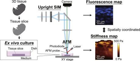
Living tissues each exhibit a distinct stiffness, which provides cells with key environmental cues that regulate their behaviors. Despite this significance, our understanding of the spatiotemporal dynamics and the biological roles of stiffness in three-dimensional tissues is currently limited due to a lack of appropriate measurement techniques. To address this issue, we propose a new method combining upright structured illumination microscopy (USIM) and atomic force microscopy (AFM) to obtain precisely coordinated stiffness maps and biomolecular fluorescence images of thick living tissue slices. Using mouse embryonic and adult skin as a representative tissue with mechanically heterogeneous structures inside, we validate the measurement principle of USIM-AFM. Live measurement of tissue stiffness distributions revealed the highly heterogeneous mechanical nature of skin, including nucleated/enucleated epithelium, mesenchyme, and hair follicle, as well as the role of collagens in maintaining its integrity. Furthermore, quantitative analysis comparing stiffness distributions in live tissue samples with those in preserved tissues, including formalin-fixed and cryopreserved tissue samples, unveiled the distinct impacts of preservation processes on tissue stiffness patterns. This series of experiments highlights the importance of live mechanical testing of tissue-scale samples to accurately capture the true spatiotemporal variations in mechanical properties. Our USIM-AFM technique provides a new methodology to reveal the dynamic nature of tissue stiffness and its correlation with biomolecular distributions in live tissues and thus could serve as a technical basis for exploring tissue-scale mechanobiology.
Statement of significance
Stiffness, a simple mechanical parameter, has drawn attention in understanding the mechanobiological principles underlying the homeostasis and pathology of living tissues. To explore tissue-scale mechanobiology, we propose a technique integrating an upright structured illumination microscope and an atomic force microscope. This technique enables live measurements of stiffness distribution and fluorescent observation of thick living tissue slices. Experiments revealed the highly heterogeneous mechanical nature of mouse embryonic and adult skin in three dimensions and the previously unnoticed influences of preservation techniques on the mechanical properties of tissue at microscopic resolution. This study provides a new technical platform for live stiffness measurement and biomolecular observation of tissue-scale samples with micron-scale resolution, thus contributing to future studies of tissue- and organ-scale mechanobiology.
期刊介绍:
Acta Biomaterialia is a monthly peer-reviewed scientific journal published by Elsevier. The journal was established in January 2005. The editor-in-chief is W.R. Wagner (University of Pittsburgh). The journal covers research in biomaterials science, including the interrelationship of biomaterial structure and function from macroscale to nanoscale. Topical coverage includes biomedical and biocompatible materials.