一只雄性弯刀角大羚羊(Oryx dammah)严重角感染的计算机断层扫描特征。

IF 1.3 2区 农林科学 Q2 VETERINARY SCIENCES
Kayla Le, Victoria Riggs, Seng Wai Yap, Martina Ernestova, Kimberley Sebastian
{"title":"一只雄性弯刀角大羚羊(Oryx dammah)严重角感染的计算机断层扫描特征。","authors":"Kayla Le, Victoria Riggs, Seng Wai Yap, Martina Ernestova, Kimberley Sebastian","doi":"10.1111/vru.13440","DOIUrl":null,"url":null,"abstract":"<p><p>A 3-year-old male Scimitar-horned oryx presented for evaluation of an acutely deformed right horn with right head tilt and right facial pain. Computed tomographic evaluation revealed an increased volume of central fluid/soft tissue attenuation with gas-attenuating foci within the right horn. The right horn was amputated at the right horn base. Imaging and histopathologic features were consistent with emphysematous osteomyelitis. Following treatment, the patient returned to normal behavior. This is the first veterinary report describing the computed tomographic features of a horn infection in a Scimitar-horned oryx.</p>","PeriodicalId":23581,"journal":{"name":"Veterinary Radiology & Ultrasound","volume":null,"pages":null},"PeriodicalIF":1.3000,"publicationDate":"2024-10-09","publicationTypes":"Journal Article","fieldsOfStudy":null,"isOpenAccess":false,"openAccessPdf":"","citationCount":"0","resultStr":"{\"title\":\"Computed tomographic features of severe horn infection in a male Scimitar-horned oryx (Oryx dammah).\",\"authors\":\"Kayla Le, Victoria Riggs, Seng Wai Yap, Martina Ernestova, Kimberley Sebastian\",\"doi\":\"10.1111/vru.13440\",\"DOIUrl\":null,\"url\":null,\"abstract\":\"<p><p>A 3-year-old male Scimitar-horned oryx presented for evaluation of an acutely deformed right horn with right head tilt and right facial pain. Computed tomographic evaluation revealed an increased volume of central fluid/soft tissue attenuation with gas-attenuating foci within the right horn. The right horn was amputated at the right horn base. Imaging and histopathologic features were consistent with emphysematous osteomyelitis. Following treatment, the patient returned to normal behavior. This is the first veterinary report describing the computed tomographic features of a horn infection in a Scimitar-horned oryx.</p>\",\"PeriodicalId\":23581,\"journal\":{\"name\":\"Veterinary Radiology & Ultrasound\",\"volume\":null,\"pages\":null},\"PeriodicalIF\":1.3000,\"publicationDate\":\"2024-10-09\",\"publicationTypes\":\"Journal Article\",\"fieldsOfStudy\":null,\"isOpenAccess\":false,\"openAccessPdf\":\"\",\"citationCount\":\"0\",\"resultStr\":null,\"platform\":\"Semanticscholar\",\"paperid\":null,\"PeriodicalName\":\"Veterinary Radiology & Ultrasound\",\"FirstCategoryId\":\"97\",\"ListUrlMain\":\"https://doi.org/10.1111/vru.13440\",\"RegionNum\":2,\"RegionCategory\":\"农林科学\",\"ArticlePicture\":[],\"TitleCN\":null,\"AbstractTextCN\":null,\"PMCID\":null,\"EPubDate\":\"\",\"PubModel\":\"\",\"JCR\":\"Q2\",\"JCRName\":\"VETERINARY SCIENCES\",\"Score\":null,\"Total\":0}","platform":"Semanticscholar","paperid":null,"PeriodicalName":"Veterinary Radiology & Ultrasound","FirstCategoryId":"97","ListUrlMain":"https://doi.org/10.1111/vru.13440","RegionNum":2,"RegionCategory":"农林科学","ArticlePicture":[],"TitleCN":null,"AbstractTextCN":null,"PMCID":null,"EPubDate":"","PubModel":"","JCR":"Q2","JCRName":"VETERINARY SCIENCES","Score":null,"Total":0}
引用次数: 0

摘要

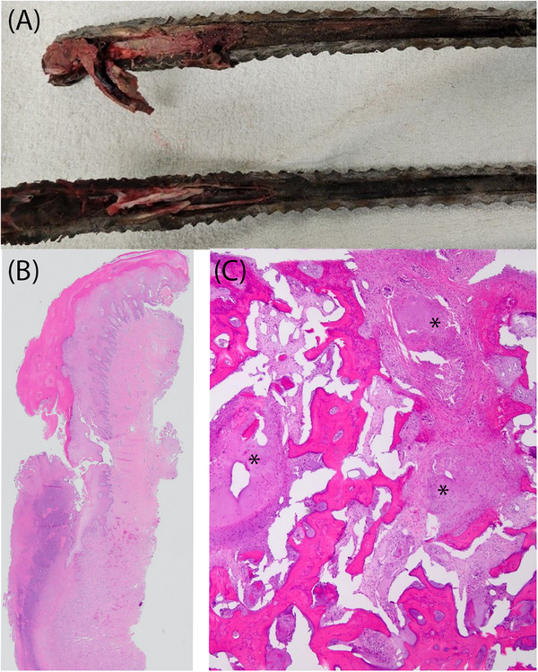
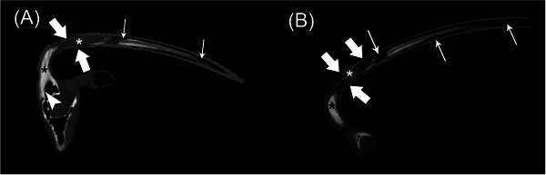
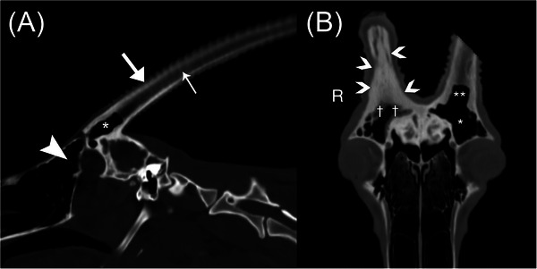
一只 3 岁的雄性弯刀角大羚羊因右侧角急性畸形、右头倾斜和右面部疼痛前来就诊。计算机断层扫描评估显示,右角中央液体/软组织衰减体积增大,并伴有气体衰减灶。右侧角被从右侧角基部截除。影像学和组织病理学特征与气肿性骨髓炎一致。治疗后,患者恢复了正常行为。这是第一份描述弯刀角大羚羊角感染计算机断层扫描特征的兽医报告。
本文章由计算机程序翻译,如有差异,请以英文原文为准。
Computed tomographic features of severe horn infection in a male Scimitar-horned oryx (Oryx dammah).

A 3-year-old male Scimitar-horned oryx presented for evaluation of an acutely deformed right horn with right head tilt and right facial pain. Computed tomographic evaluation revealed an increased volume of central fluid/soft tissue attenuation with gas-attenuating foci within the right horn. The right horn was amputated at the right horn base. Imaging and histopathologic features were consistent with emphysematous osteomyelitis. Following treatment, the patient returned to normal behavior. This is the first veterinary report describing the computed tomographic features of a horn infection in a Scimitar-horned oryx.

求助全文
通过发布文献求助,成功后即可免费获取论文全文。 去求助
来源期刊
Veterinary Radiology & Ultrasound
Veterinary Radiology & Ultrasound 农林科学-兽医学
CiteScore
2.40
自引率
17.60%
发文量
133
审稿时长
8-16 weeks
期刊介绍: Veterinary Radiology & Ultrasound is a bimonthly, international, peer-reviewed, research journal devoted to the fields of veterinary diagnostic imaging and radiation oncology. Established in 1958, it is owned by the American College of Veterinary Radiology and is also the official journal for six affiliate veterinary organizations. Veterinary Radiology & Ultrasound is represented on the International Committee of Medical Journal Editors, World Association of Medical Editors, and Committee on Publication Ethics. The mission of Veterinary Radiology & Ultrasound is to serve as a leading resource for high quality articles that advance scientific knowledge and standards of clinical practice in the areas of veterinary diagnostic radiology, computed tomography, magnetic resonance imaging, ultrasonography, nuclear imaging, radiation oncology, and interventional radiology. Manuscript types include original investigations, imaging diagnosis reports, review articles, editorials and letters to the Editor. Acceptance criteria include originality, significance, quality, reader interest, composition and adherence to author guidelines.
×
引用
GB/T 7714-2015
复制
MLA
复制
APA
复制
导出至
BibTeX EndNote RefMan NoteFirst NoteExpress
×
提示
您的信息不完整,为了账户安全,请先补充。
现在去补充
×
提示
您因"违规操作"
具体请查看互助需知
我知道了
×
提示
确定
请完成安全验证×
copy
已复制链接
快去分享给好友吧!
我知道了
右上角分享
点击右上角分享
0
联系我们:info@booksci.cn Book学术提供免费学术资源搜索服务,方便国内外学者检索中英文文献。致力于提供最便捷和优质的服务体验。 Copyright © 2023 布克学术 All rights reserved.
京ICP备2023020795号-1
ghs 京公网安备 11010802042870号
Book学术文献互助
Book学术文献互助群
群 号:481959085
Book学术官方微信