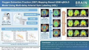
利用多延迟 PCASL,结合定量易感性图谱和定量血氧饱和度依赖性成像模型,绘制氧萃取分数图。

IF 2.7 4区 医学 Q3 NEUROSCIENCES
Xiaoyi Liu , Yayan Yin , Yi Shan , Wang Chao , Jingkai Li , Yue Zhang , Qiongge Li , Jing Liu , Jie Lu
{"title":"利用多延迟 PCASL,结合定量易感性图谱和定量血氧饱和度依赖性成像模型,绘制氧萃取分数图。","authors":"Xiaoyi Liu ,&nbsp;Yayan Yin ,&nbsp;Yi Shan ,&nbsp;Wang Chao ,&nbsp;Jingkai Li ,&nbsp;Yue Zhang ,&nbsp;Qiongge Li ,&nbsp;Jing Liu ,&nbsp;Jie Lu","doi":"10.1016/j.brainres.2024.149259","DOIUrl":null,"url":null,"abstract":"<div><h3>Background and purpose</h3><div>The oxygen extraction fraction is an essential biomarker for the assessment of brain metabolism. A recently proposed method combined with quantitative susceptibility mapping and quantitative blood oxygen level-dependent magnitude enables noninvasive mapping of the oxygen extraction fraction. Our study investigated the oxygen extraction fraction mapping variations of single-delay and multi-delay arterial spin-labeling.</div></div><div><h3>Materials and methods</h3><div>A total of twenty healthy participants were enrolled. The multi-echo spoiled gradient-echo, multi-delay arterial spin-labeling, and magnetization-prepared rapid gradient echo sequences were acquired at 3.0 T. The mean oxygen extraction fraction was generated under a single delay time of 1780 ms, multi-delay arterial spin-labeling of transit-corrected cerebral blood flow, and multi-delay arterial spin-labeling of arterial cerebral blood volume. The results were compared via paired t tests and the Wilcoxon test. Linear regression analyses were used to investigate the relationships among the oxygen extraction fraction, cerebral blood flow, and venous cerebral blood volume.</div></div><div><h3>Results</h3><div>The oxygen extraction fraction estimate with multi-delay arterial spin-labeling yielded a significantly lower value than that with single-delay arterial spin-labeling. The average values for the whole brain under single-delay arterial spin-labeling, multi-delay arterial spin-labeling of transit-corrected cerebral blood flow, and multi-delay arterial spin-labeling of arterial cerebral blood volume were 41.5 ± 1.7 % (P &lt; 0.05), 41.3 ± 1.9 % (P &lt; 0.001), and 40.9 ± 1.9 % (N = 20), respectively. The oxygen extraction fraction also showed a significant inverse correlation with the venous cerebral blood volume under steady-state conditions when multi-delay arterial spin-labeling was used (r = 0.5834, p = 0.0069).</div></div><div><h3>Conclusion</h3><div>These findings suggest that the oxygen extraction fraction is significantly impacted by the arterial spin-labeling methods used in the quantitative susceptibility mapping plus the quantitative blood oxygen level-dependent model, indicating that the differences should be accounted for when employing oxygen extraction fraction mapping based on this model in diseases.</div></div>","PeriodicalId":9083,"journal":{"name":"Brain Research","volume":"1846 ","pages":"Article 149259"},"PeriodicalIF":2.7000,"publicationDate":"2024-10-03","publicationTypes":"Journal Article","fieldsOfStudy":null,"isOpenAccess":false,"openAccessPdf":"","citationCount":"0","resultStr":"{\"title\":\"Oxygen extraction fraction mapping based combining quantitative susceptibility mapping and quantitative blood oxygenation level-dependent imaging model using multi-delay PCASL\",\"authors\":\"Xiaoyi Liu ,&nbsp;Yayan Yin ,&nbsp;Yi Shan ,&nbsp;Wang Chao ,&nbsp;Jingkai Li ,&nbsp;Yue Zhang ,&nbsp;Qiongge Li ,&nbsp;Jing Liu ,&nbsp;Jie Lu\",\"doi\":\"10.1016/j.brainres.2024.149259\",\"DOIUrl\":null,\"url\":null,\"abstract\":\"<div><h3>Background and purpose</h3><div>The oxygen extraction fraction is an essential biomarker for the assessment of brain metabolism. A recently proposed method combined with quantitative susceptibility mapping and quantitative blood oxygen level-dependent magnitude enables noninvasive mapping of the oxygen extraction fraction. Our study investigated the oxygen extraction fraction mapping variations of single-delay and multi-delay arterial spin-labeling.</div></div><div><h3>Materials and methods</h3><div>A total of twenty healthy participants were enrolled. The multi-echo spoiled gradient-echo, multi-delay arterial spin-labeling, and magnetization-prepared rapid gradient echo sequences were acquired at 3.0 T. The mean oxygen extraction fraction was generated under a single delay time of 1780 ms, multi-delay arterial spin-labeling of transit-corrected cerebral blood flow, and multi-delay arterial spin-labeling of arterial cerebral blood volume. The results were compared via paired t tests and the Wilcoxon test. Linear regression analyses were used to investigate the relationships among the oxygen extraction fraction, cerebral blood flow, and venous cerebral blood volume.</div></div><div><h3>Results</h3><div>The oxygen extraction fraction estimate with multi-delay arterial spin-labeling yielded a significantly lower value than that with single-delay arterial spin-labeling. The average values for the whole brain under single-delay arterial spin-labeling, multi-delay arterial spin-labeling of transit-corrected cerebral blood flow, and multi-delay arterial spin-labeling of arterial cerebral blood volume were 41.5 ± 1.7 % (P &lt; 0.05), 41.3 ± 1.9 % (P &lt; 0.001), and 40.9 ± 1.9 % (N = 20), respectively. The oxygen extraction fraction also showed a significant inverse correlation with the venous cerebral blood volume under steady-state conditions when multi-delay arterial spin-labeling was used (r = 0.5834, p = 0.0069).</div></div><div><h3>Conclusion</h3><div>These findings suggest that the oxygen extraction fraction is significantly impacted by the arterial spin-labeling methods used in the quantitative susceptibility mapping plus the quantitative blood oxygen level-dependent model, indicating that the differences should be accounted for when employing oxygen extraction fraction mapping based on this model in diseases.</div></div>\",\"PeriodicalId\":9083,\"journal\":{\"name\":\"Brain Research\",\"volume\":\"1846 \",\"pages\":\"Article 149259\"},\"PeriodicalIF\":2.7000,\"publicationDate\":\"2024-10-03\",\"publicationTypes\":\"Journal Article\",\"fieldsOfStudy\":null,\"isOpenAccess\":false,\"openAccessPdf\":\"\",\"citationCount\":\"0\",\"resultStr\":null,\"platform\":\"Semanticscholar\",\"paperid\":null,\"PeriodicalName\":\"Brain Research\",\"FirstCategoryId\":\"3\",\"ListUrlMain\":\"https://www.sciencedirect.com/science/article/pii/S0006899324005134\",\"RegionNum\":4,\"RegionCategory\":\"医学\",\"ArticlePicture\":[],\"TitleCN\":null,\"AbstractTextCN\":null,\"PMCID\":null,\"EPubDate\":\"\",\"PubModel\":\"\",\"JCR\":\"Q3\",\"JCRName\":\"NEUROSCIENCES\",\"Score\":null,\"Total\":0}","platform":"Semanticscholar","paperid":null,"PeriodicalName":"Brain Research","FirstCategoryId":"3","ListUrlMain":"https://www.sciencedirect.com/science/article/pii/S0006899324005134","RegionNum":4,"RegionCategory":"医学","ArticlePicture":[],"TitleCN":null,"AbstractTextCN":null,"PMCID":null,"EPubDate":"","PubModel":"","JCR":"Q3","JCRName":"NEUROSCIENCES","Score":null,"Total":0}
引用次数: 0

摘要

背景和目的:氧萃取分数是评估大脑新陈代谢的重要生物标志物。最近提出的一种方法结合了定量易感性图谱和定量血氧水平相关幅度,可以无创地绘制氧萃取分数图谱。我们的研究调查了单延迟和多延迟动脉自旋标记的氧萃取分数绘图变化:材料和方法:共招募了 20 名健康参与者。在 3.0 T 下采集了多回波破坏梯度回波、多延迟动脉自旋标记和磁化准备快速梯度回波序列。在 1780 毫秒的单一延迟时间、多延迟动脉自旋标记的过境校正脑血流和多延迟动脉自旋标记的动脉脑血量条件下生成平均析氧分数。结果通过配对 t 检验和 Wilcoxon 检验进行比较。线性回归分析用于研究氧提取率、脑血流量和静脉脑血量之间的关系:结果:用多延迟动脉自旋标记法估算的氧萃取分数明显低于用单延迟动脉自旋标记法估算的氧萃取分数。单延迟动脉自旋标记法、多延迟动脉自旋标记法的过境校正脑血流量和多延迟动脉自旋标记法的动脉脑血量的全脑平均值分别为 41.5 ± 1.7 %(P 结论:多延迟动脉自旋标记法和单延迟动脉自旋标记法的全脑平均值均低于单延迟动脉自旋标记法:这些研究结果表明,氧萃取分数受定量易感性图谱加定量血氧水平依赖模型中使用的动脉自旋标记方法的显著影响,这表明在疾病中使用基于该模型的氧萃取分数图谱时应考虑这些差异。
本文章由计算机程序翻译,如有差异,请以英文原文为准。

Oxygen extraction fraction mapping based combining quantitative susceptibility mapping and quantitative blood oxygenation level-dependent imaging model using multi-delay PCASL

Oxygen extraction fraction mapping based combining quantitative susceptibility mapping and quantitative blood oxygenation level-dependent imaging model using multi-delay PCASL

Background and purpose

The oxygen extraction fraction is an essential biomarker for the assessment of brain metabolism. A recently proposed method combined with quantitative susceptibility mapping and quantitative blood oxygen level-dependent magnitude enables noninvasive mapping of the oxygen extraction fraction. Our study investigated the oxygen extraction fraction mapping variations of single-delay and multi-delay arterial spin-labeling.

Materials and methods

A total of twenty healthy participants were enrolled. The multi-echo spoiled gradient-echo, multi-delay arterial spin-labeling, and magnetization-prepared rapid gradient echo sequences were acquired at 3.0 T. The mean oxygen extraction fraction was generated under a single delay time of 1780 ms, multi-delay arterial spin-labeling of transit-corrected cerebral blood flow, and multi-delay arterial spin-labeling of arterial cerebral blood volume. The results were compared via paired t tests and the Wilcoxon test. Linear regression analyses were used to investigate the relationships among the oxygen extraction fraction, cerebral blood flow, and venous cerebral blood volume.

Results

The oxygen extraction fraction estimate with multi-delay arterial spin-labeling yielded a significantly lower value than that with single-delay arterial spin-labeling. The average values for the whole brain under single-delay arterial spin-labeling, multi-delay arterial spin-labeling of transit-corrected cerebral blood flow, and multi-delay arterial spin-labeling of arterial cerebral blood volume were 41.5 ± 1.7 % (P < 0.05), 41.3 ± 1.9 % (P < 0.001), and 40.9 ± 1.9 % (N = 20), respectively. The oxygen extraction fraction also showed a significant inverse correlation with the venous cerebral blood volume under steady-state conditions when multi-delay arterial spin-labeling was used (r = 0.5834, p = 0.0069).

Conclusion

These findings suggest that the oxygen extraction fraction is significantly impacted by the arterial spin-labeling methods used in the quantitative susceptibility mapping plus the quantitative blood oxygen level-dependent model, indicating that the differences should be accounted for when employing oxygen extraction fraction mapping based on this model in diseases.
求助全文
通过发布文献求助,成功后即可免费获取论文全文。 去求助
来源期刊
Brain Research
Brain Research 医学-神经科学
CiteScore
5.90
自引率
3.40%
发文量
268
审稿时长
47 days
期刊介绍: An international multidisciplinary journal devoted to fundamental research in the brain sciences. Brain Research publishes papers reporting interdisciplinary investigations of nervous system structure and function that are of general interest to the international community of neuroscientists. As is evident from the journals name, its scope is broad, ranging from cellular and molecular studies through systems neuroscience, cognition and disease. Invited reviews are also published; suggestions for and inquiries about potential reviews are welcomed. With the appearance of the final issue of the 2011 subscription, Vol. 67/1-2 (24 June 2011), Brain Research Reviews has ceased publication as a distinct journal separate from Brain Research. Review articles accepted for Brain Research are now published in that journal.
×
引用
GB/T 7714-2015
复制
MLA
复制
APA
复制
导出至
BibTeX EndNote RefMan NoteFirst NoteExpress
×
提示
您的信息不完整,为了账户安全,请先补充。
现在去补充
×
提示
您因"违规操作"
具体请查看互助需知
我知道了
×
提示
确定
请完成安全验证×
copy
已复制链接
快去分享给好友吧!
我知道了
右上角分享
点击右上角分享
0
联系我们:info@booksci.cn Book学术提供免费学术资源搜索服务,方便国内外学者检索中英文文献。致力于提供最便捷和优质的服务体验。 Copyright © 2023 布克学术 All rights reserved.
京ICP备2023020795号-1
ghs 京公网安备 11010802042870号
Book学术文献互助
Book学术文献互助群
群 号:481959085
Book学术官方微信