{"title":"利用双参数磁共振成像的机器学习模型区分有临床意义和无临床意义的前列腺癌。","authors":"Hakan Ayyıldız, Okan İnce, Esin Korkut, Merve Gülbiz Dağoğlu Kartal, Atadan Tunacı, Şükrü Mehmet Ertürk","doi":"10.4274/dir.2024.242856","DOIUrl":null,"url":null,"abstract":"<p><strong>Purpose: </strong>This study aims to demonstrate the performance of machine learning algorithms to distinguish clinically significant prostate cancer (csPCa) from clinically insignificant prostate cancer (ciPCa) in prostate bi-parametric magnetic resonance imaging (MRI) using radiomics features.</p><p><strong>Methods: </strong>MRI images of patients who were diagnosed with cancer with histopathological confirmation following prostate MRI were collected retrospectively. Patients with a Gleason score of 3+3 were considered to have clinically ciPCa, and patients with a Gleason score of 3+4 and above were considered to have csPCa. Radiomics features were extracted from T2-weighted (T2W) images, apparent diffusion coefficient (ADC) images, and their corresponding Laplacian of Gaussian (LoG) filtered versions. Additionally, a third feature subset was created by combining the T2W and ADC images, enhancing the analysis with an integrated approach. Once the features were extracted, Pearson’s correlation coefficient and selection were performed using wrapper-based sequential algorithms. The models were then built using support vector machine (SVM) and logistic regression (LR) machine learning algorithms. The models were validated using a five-fold cross-validation technique.</p><p><strong>Results: </strong>This study included 77 patients, 30 with ciPCA and 47 with csPCA. From each image, four images were extracted with LoG filtering, and 111 features were obtained from each image. After feature selection, 5 features were obtained from T2W images, 5 from ADC images, and 15 from the combined dataset. In the SVM model, area under the curve (AUC) values of 0.64 for T2W, 0.86 for ADC, and 0.86 for the combined dataset were obtained in the test set. In the LR model, AUC values of 0.79 for T2W, 0.86 for ADC, and 0.85 for the combined dataset were obtained.</p><p><strong>Conclusion: </strong>Machine learning models developed with radiomics can provide a decision support system to complement pathology results and help avoid invasive procedures such as re-biopsies or follow-up biopsies that are sometimes necessary today.</p><p><strong>Clinical significance: </strong>This study demonstrates that machine learning models using radiomics features derived from bi-parametric MRI can discriminate csPCa from clinically insignificant PCa. These findings suggest that radiomics-based machine learning models have the potential to reduce the need for re-biopsy in cases of indeterminate pathology, assist in diagnosing pathology–radiology discordance, and support treatment decision-making in the management of PCa.</p>","PeriodicalId":11341,"journal":{"name":"Diagnostic and interventional radiology","volume":" ","pages":"313-320"},"PeriodicalIF":1.7000,"publicationDate":"2025-07-08","publicationTypes":"Journal Article","fieldsOfStudy":null,"isOpenAccess":false,"openAccessPdf":"https://www.ncbi.nlm.nih.gov/pmc/articles/PMC12239529/pdf/","citationCount":"0","resultStr":"{\"title\":\"Machine learning models for discriminating clinically significant from clinically insignificant prostate cancer using bi-parametric magnetic resonance imaging\",\"authors\":\"Hakan Ayyıldız, Okan İnce, Esin Korkut, Merve Gülbiz Dağoğlu Kartal, Atadan Tunacı, Şükrü Mehmet Ertürk\",\"doi\":\"10.4274/dir.2024.242856\",\"DOIUrl\":null,\"url\":null,\"abstract\":\"<p><strong>Purpose: </strong>This study aims to demonstrate the performance of machine learning algorithms to distinguish clinically significant prostate cancer (csPCa) from clinically insignificant prostate cancer (ciPCa) in prostate bi-parametric magnetic resonance imaging (MRI) using radiomics features.</p><p><strong>Methods: </strong>MRI images of patients who were diagnosed with cancer with histopathological confirmation following prostate MRI were collected retrospectively. Patients with a Gleason score of 3+3 were considered to have clinically ciPCa, and patients with a Gleason score of 3+4 and above were considered to have csPCa. Radiomics features were extracted from T2-weighted (T2W) images, apparent diffusion coefficient (ADC) images, and their corresponding Laplacian of Gaussian (LoG) filtered versions. Additionally, a third feature subset was created by combining the T2W and ADC images, enhancing the analysis with an integrated approach. Once the features were extracted, Pearson’s correlation coefficient and selection were performed using wrapper-based sequential algorithms. The models were then built using support vector machine (SVM) and logistic regression (LR) machine learning algorithms. The models were validated using a five-fold cross-validation technique.</p><p><strong>Results: </strong>This study included 77 patients, 30 with ciPCA and 47 with csPCA. From each image, four images were extracted with LoG filtering, and 111 features were obtained from each image. After feature selection, 5 features were obtained from T2W images, 5 from ADC images, and 15 from the combined dataset. In the SVM model, area under the curve (AUC) values of 0.64 for T2W, 0.86 for ADC, and 0.86 for the combined dataset were obtained in the test set. In the LR model, AUC values of 0.79 for T2W, 0.86 for ADC, and 0.85 for the combined dataset were obtained.</p><p><strong>Conclusion: </strong>Machine learning models developed with radiomics can provide a decision support system to complement pathology results and help avoid invasive procedures such as re-biopsies or follow-up biopsies that are sometimes necessary today.</p><p><strong>Clinical significance: </strong>This study demonstrates that machine learning models using radiomics features derived from bi-parametric MRI can discriminate csPCa from clinically insignificant PCa. These findings suggest that radiomics-based machine learning models have the potential to reduce the need for re-biopsy in cases of indeterminate pathology, assist in diagnosing pathology–radiology discordance, and support treatment decision-making in the management of PCa.</p>\",\"PeriodicalId\":11341,\"journal\":{\"name\":\"Diagnostic and interventional radiology\",\"volume\":\" \",\"pages\":\"313-320\"},\"PeriodicalIF\":1.7000,\"publicationDate\":\"2025-07-08\",\"publicationTypes\":\"Journal Article\",\"fieldsOfStudy\":null,\"isOpenAccess\":false,\"openAccessPdf\":\"https://www.ncbi.nlm.nih.gov/pmc/articles/PMC12239529/pdf/\",\"citationCount\":\"0\",\"resultStr\":null,\"platform\":\"Semanticscholar\",\"paperid\":null,\"PeriodicalName\":\"Diagnostic and interventional radiology\",\"FirstCategoryId\":\"3\",\"ListUrlMain\":\"https://doi.org/10.4274/dir.2024.242856\",\"RegionNum\":4,\"RegionCategory\":\"医学\",\"ArticlePicture\":[],\"TitleCN\":null,\"AbstractTextCN\":null,\"PMCID\":null,\"EPubDate\":\"2024/10/1 0:00:00\",\"PubModel\":\"Epub\",\"JCR\":\"Q3\",\"JCRName\":\"RADIOLOGY, NUCLEAR MEDICINE & MEDICAL IMAGING\",\"Score\":null,\"Total\":0}","platform":"Semanticscholar","paperid":null,"PeriodicalName":"Diagnostic and interventional radiology","FirstCategoryId":"3","ListUrlMain":"https://doi.org/10.4274/dir.2024.242856","RegionNum":4,"RegionCategory":"医学","ArticlePicture":[],"TitleCN":null,"AbstractTextCN":null,"PMCID":null,"EPubDate":"2024/10/1 0:00:00","PubModel":"Epub","JCR":"Q3","JCRName":"RADIOLOGY, NUCLEAR MEDICINE & MEDICAL IMAGING","Score":null,"Total":0}
Machine learning models for discriminating clinically significant from clinically insignificant prostate cancer using bi-parametric magnetic resonance imaging
Purpose: This study aims to demonstrate the performance of machine learning algorithms to distinguish clinically significant prostate cancer (csPCa) from clinically insignificant prostate cancer (ciPCa) in prostate bi-parametric magnetic resonance imaging (MRI) using radiomics features.
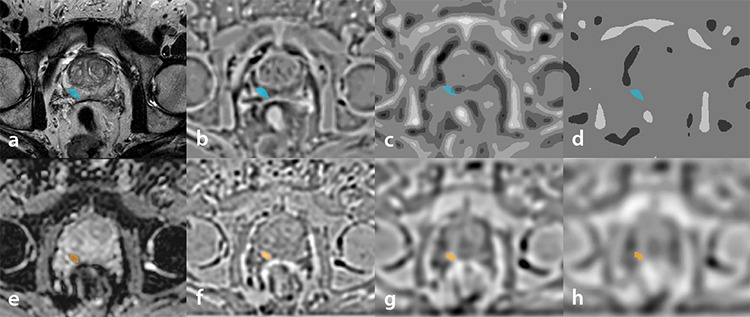
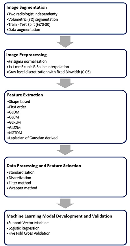
Methods: MRI images of patients who were diagnosed with cancer with histopathological confirmation following prostate MRI were collected retrospectively. Patients with a Gleason score of 3+3 were considered to have clinically ciPCa, and patients with a Gleason score of 3+4 and above were considered to have csPCa. Radiomics features were extracted from T2-weighted (T2W) images, apparent diffusion coefficient (ADC) images, and their corresponding Laplacian of Gaussian (LoG) filtered versions. Additionally, a third feature subset was created by combining the T2W and ADC images, enhancing the analysis with an integrated approach. Once the features were extracted, Pearson’s correlation coefficient and selection were performed using wrapper-based sequential algorithms. The models were then built using support vector machine (SVM) and logistic regression (LR) machine learning algorithms. The models were validated using a five-fold cross-validation technique.
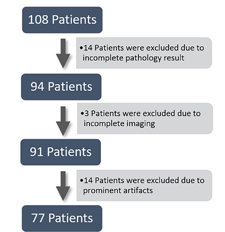
Results: This study included 77 patients, 30 with ciPCA and 47 with csPCA. From each image, four images were extracted with LoG filtering, and 111 features were obtained from each image. After feature selection, 5 features were obtained from T2W images, 5 from ADC images, and 15 from the combined dataset. In the SVM model, area under the curve (AUC) values of 0.64 for T2W, 0.86 for ADC, and 0.86 for the combined dataset were obtained in the test set. In the LR model, AUC values of 0.79 for T2W, 0.86 for ADC, and 0.85 for the combined dataset were obtained.
Conclusion: Machine learning models developed with radiomics can provide a decision support system to complement pathology results and help avoid invasive procedures such as re-biopsies or follow-up biopsies that are sometimes necessary today.
Clinical significance: This study demonstrates that machine learning models using radiomics features derived from bi-parametric MRI can discriminate csPCa from clinically insignificant PCa. These findings suggest that radiomics-based machine learning models have the potential to reduce the need for re-biopsy in cases of indeterminate pathology, assist in diagnosing pathology–radiology discordance, and support treatment decision-making in the management of PCa.
期刊介绍:
Diagnostic and Interventional Radiology (Diagn Interv Radiol) is the open access, online-only official publication of Turkish Society of Radiology. It is published bimonthly and the journal’s publication language is English.
The journal is a medium for original articles, reviews, pictorial essays, technical notes related to all fields of diagnostic and interventional radiology.

 求助内容:
求助内容: 应助结果提醒方式:


