Rong Yang, Lee Mui Lee, YaoMin Zhu, Wen Yuan Jia, Wei Yao, Yue Yu, Shu Jun Wu
{"title":"颞下颌关节椎间盘穿孔与关节退行性病变之间的相关性:CBCT 和临床分析","authors":"Rong Yang, Lee Mui Lee, YaoMin Zhu, Wen Yuan Jia, Wei Yao, Yue Yu, Shu Jun Wu","doi":"10.1111/joor.13866","DOIUrl":null,"url":null,"abstract":"<div>\n \n \n <section>\n \n <h3> Objectives</h3>\n \n <p>To analyse the correlation between temporomandibular joint (TMJ) disc perforation and degenerative joint changes (DJC) on cone-beam computed tomography (CBCT) and related factors.</p>\n </section>\n \n <section>\n \n <h3> Study Design</h3>\n \n <p>A total of 238 female patients with anterior disc displacement without reduction (ADDwoR), accounting for 348 affected joints, requiring TMJ disc open anchorage surgery were included in the study conducted from June 2021 to August 2022. Following TMJ disc open anchorage surgery, patients were divided into two groups: disc perforation (DP) and disc non-perforation (DNP). CBCT was utilised to assess different grades of condyle and articular eminence degenerative changes, and comparisons were made between the two groups regarding DJC and clinically relevant factors.</p>\n </section>\n \n <section>\n \n <h3> Results</h3>\n \n <p>In comparison with the DNP group, the DP group exhibited statistically significant differences in mid- and late-stage condyle bone degenerative changes and bone alterations of the articular eminence (odds ratio [OR] = 7.822; 95% CI [4.438–13.785]; <i>p</i> < 0.001 and OR = 5.575; 95% CI [3.128–9.936]; <i>p</i> < 0.001). Additionally, persistent joint sounds (OR = 1.932; 95% CI [1.011–3.691]; <i>p</i> = 0.046) and longer disease duration (OR = 4.901; 95% CI [2.395–10.028]; <i>p</i> < 0.001) demonstrated statistically significant differences. However, no significant differences were observed between the two groups in terms of age, joint pain and limited mouth opening.</p>\n </section>\n \n <section>\n \n <h3> Conclusions</h3>\n \n <p>Bone degeneration changes in TMJ CBCT images are a high possible risk factor for DP. With an escalation in the degree of condyle degeneration, the risk of DP may increased correspondingly. Persistent joint sounds and extended duration of the disease were also confirmed to be noteworthy clinical risks of DP.</p>\n </section>\n </div>","PeriodicalId":16605,"journal":{"name":"Journal of oral rehabilitation","volume":"51 12","pages":"2675-2682"},"PeriodicalIF":3.1000,"publicationDate":"2024-09-27","publicationTypes":"Journal Article","fieldsOfStudy":null,"isOpenAccess":false,"openAccessPdf":"","citationCount":"0","resultStr":"{\"title\":\"Correlation Between Temporomandibular Joint Disc Perforation and Degenerative Joint Changes: A CBCT and Clinical Analysis\",\"authors\":\"Rong Yang, Lee Mui Lee, YaoMin Zhu, Wen Yuan Jia, Wei Yao, Yue Yu, Shu Jun Wu\",\"doi\":\"10.1111/joor.13866\",\"DOIUrl\":null,\"url\":null,\"abstract\":\"<div>\\n \\n \\n <section>\\n \\n <h3> Objectives</h3>\\n \\n <p>To analyse the correlation between temporomandibular joint (TMJ) disc perforation and degenerative joint changes (DJC) on cone-beam computed tomography (CBCT) and related factors.</p>\\n </section>\\n \\n <section>\\n \\n <h3> Study Design</h3>\\n \\n <p>A total of 238 female patients with anterior disc displacement without reduction (ADDwoR), accounting for 348 affected joints, requiring TMJ disc open anchorage surgery were included in the study conducted from June 2021 to August 2022. Following TMJ disc open anchorage surgery, patients were divided into two groups: disc perforation (DP) and disc non-perforation (DNP). CBCT was utilised to assess different grades of condyle and articular eminence degenerative changes, and comparisons were made between the two groups regarding DJC and clinically relevant factors.</p>\\n </section>\\n \\n <section>\\n \\n <h3> Results</h3>\\n \\n <p>In comparison with the DNP group, the DP group exhibited statistically significant differences in mid- and late-stage condyle bone degenerative changes and bone alterations of the articular eminence (odds ratio [OR] = 7.822; 95% CI [4.438–13.785]; <i>p</i> < 0.001 and OR = 5.575; 95% CI [3.128–9.936]; <i>p</i> < 0.001). Additionally, persistent joint sounds (OR = 1.932; 95% CI [1.011–3.691]; <i>p</i> = 0.046) and longer disease duration (OR = 4.901; 95% CI [2.395–10.028]; <i>p</i> < 0.001) demonstrated statistically significant differences. However, no significant differences were observed between the two groups in terms of age, joint pain and limited mouth opening.</p>\\n </section>\\n \\n <section>\\n \\n <h3> Conclusions</h3>\\n \\n <p>Bone degeneration changes in TMJ CBCT images are a high possible risk factor for DP. With an escalation in the degree of condyle degeneration, the risk of DP may increased correspondingly. Persistent joint sounds and extended duration of the disease were also confirmed to be noteworthy clinical risks of DP.</p>\\n </section>\\n </div>\",\"PeriodicalId\":16605,\"journal\":{\"name\":\"Journal of oral rehabilitation\",\"volume\":\"51 12\",\"pages\":\"2675-2682\"},\"PeriodicalIF\":3.1000,\"publicationDate\":\"2024-09-27\",\"publicationTypes\":\"Journal Article\",\"fieldsOfStudy\":null,\"isOpenAccess\":false,\"openAccessPdf\":\"\",\"citationCount\":\"0\",\"resultStr\":null,\"platform\":\"Semanticscholar\",\"paperid\":null,\"PeriodicalName\":\"Journal of oral rehabilitation\",\"FirstCategoryId\":\"3\",\"ListUrlMain\":\"https://onlinelibrary.wiley.com/doi/10.1111/joor.13866\",\"RegionNum\":3,\"RegionCategory\":\"医学\",\"ArticlePicture\":[],\"TitleCN\":null,\"AbstractTextCN\":null,\"PMCID\":null,\"EPubDate\":\"\",\"PubModel\":\"\",\"JCR\":\"Q1\",\"JCRName\":\"DENTISTRY, ORAL SURGERY & MEDICINE\",\"Score\":null,\"Total\":0}","platform":"Semanticscholar","paperid":null,"PeriodicalName":"Journal of oral rehabilitation","FirstCategoryId":"3","ListUrlMain":"https://onlinelibrary.wiley.com/doi/10.1111/joor.13866","RegionNum":3,"RegionCategory":"医学","ArticlePicture":[],"TitleCN":null,"AbstractTextCN":null,"PMCID":null,"EPubDate":"","PubModel":"","JCR":"Q1","JCRName":"DENTISTRY, ORAL SURGERY & MEDICINE","Score":null,"Total":0}
Correlation Between Temporomandibular Joint Disc Perforation and Degenerative Joint Changes: A CBCT and Clinical Analysis
Objectives
To analyse the correlation between temporomandibular joint (TMJ) disc perforation and degenerative joint changes (DJC) on cone-beam computed tomography (CBCT) and related factors.
Study Design
A total of 238 female patients with anterior disc displacement without reduction (ADDwoR), accounting for 348 affected joints, requiring TMJ disc open anchorage surgery were included in the study conducted from June 2021 to August 2022. Following TMJ disc open anchorage surgery, patients were divided into two groups: disc perforation (DP) and disc non-perforation (DNP). CBCT was utilised to assess different grades of condyle and articular eminence degenerative changes, and comparisons were made between the two groups regarding DJC and clinically relevant factors.
Results
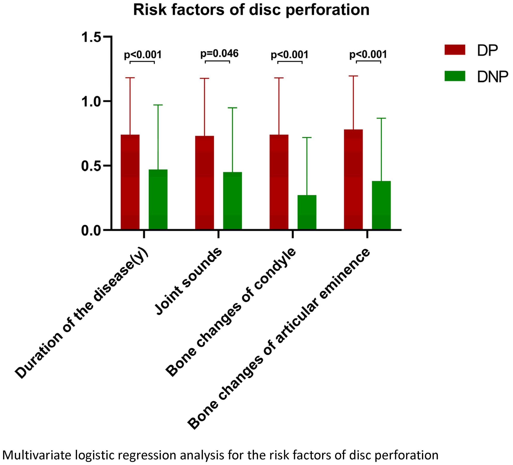
In comparison with the DNP group, the DP group exhibited statistically significant differences in mid- and late-stage condyle bone degenerative changes and bone alterations of the articular eminence (odds ratio [OR] = 7.822; 95% CI [4.438–13.785]; p < 0.001 and OR = 5.575; 95% CI [3.128–9.936]; p < 0.001). Additionally, persistent joint sounds (OR = 1.932; 95% CI [1.011–3.691]; p = 0.046) and longer disease duration (OR = 4.901; 95% CI [2.395–10.028]; p < 0.001) demonstrated statistically significant differences. However, no significant differences were observed between the two groups in terms of age, joint pain and limited mouth opening.
Conclusions
Bone degeneration changes in TMJ CBCT images are a high possible risk factor for DP. With an escalation in the degree of condyle degeneration, the risk of DP may increased correspondingly. Persistent joint sounds and extended duration of the disease were also confirmed to be noteworthy clinical risks of DP.
期刊介绍:
Journal of Oral Rehabilitation aims to be the most prestigious journal of dental research within all aspects of oral rehabilitation and applied oral physiology. It covers all diagnostic and clinical management aspects necessary to re-establish a subjective and objective harmonious oral function.
Oral rehabilitation may become necessary as a result of developmental or acquired disturbances in the orofacial region, orofacial traumas, or a variety of dental and oral diseases (primarily dental caries and periodontal diseases) and orofacial pain conditions. As such, oral rehabilitation in the twenty-first century is a matter of skilful diagnosis and minimal, appropriate intervention, the nature of which is intimately linked to a profound knowledge of oral physiology, oral biology, and dental and oral pathology.
The scientific content of the journal therefore strives to reflect the best of evidence-based clinical dentistry. Modern clinical management should be based on solid scientific evidence gathered about diagnostic procedures and the properties and efficacy of the chosen intervention (e.g. material science, biological, toxicological, pharmacological or psychological aspects). The content of the journal also reflects documentation of the possible side-effects of rehabilitation, and includes prognostic perspectives of the treatment modalities chosen.
 求助内容:
求助内容: 应助结果提醒方式:


