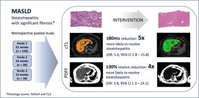
cT1 和肝脏脂肪含量的减少反映了治疗引起的 MASH 组织学改善。

IF 26.8 1区 医学 Q1 GASTROENTEROLOGY & HEPATOLOGY
Naim Alkhouri, Cayden Beyer, Elizabeth Shumbayawonda, Anneli Andersson, Kitty Yale, Timothy Rolph, Raymond T Chung, Raj Vuppalanchi, Kenneth Cusi, Rohit Loomba, Michele Pansini, Andrea Dennis
{"title":"cT1 和肝脏脂肪含量的减少反映了治疗引起的 MASH 组织学改善。","authors":"Naim Alkhouri, Cayden Beyer, Elizabeth Shumbayawonda, Anneli Andersson, Kitty Yale, Timothy Rolph, Raymond T Chung, Raj Vuppalanchi, Kenneth Cusi, Rohit Loomba, Michele Pansini, Andrea Dennis","doi":"10.1016/j.jhep.2024.08.031","DOIUrl":null,"url":null,"abstract":"<p><strong>Background & aims: </strong>MRI biomarkers of liver disease are robust and reproducible alternatives to liver biopsy. Emerging data suggest that absolute reduction in iron corrected T1 (cT1) of ≥ 80 ms and relative reduction in liver fat content of 30% reflect histological improvement. We aimed to validate the associations of changes to these noninvasive biomarkers with histological improvement, specifically the resolution of steatohepatitis.</p><p><strong>Methods: </strong>A retrospective analysis of participants from three interventional clinical trials who underwent multiparametric MRI to measure liver cT1 and liver fat content (LFC) (LiverMultiScan) alongside biopsies at baseline and end of study. Responders were defined as those achieving resolution of steatohepatitis with no worsening in fibrosis. Differences in the magnitude of change in cT1 and LFC between responders and non-responders was assessed.</p><p><strong>Results: </strong>Individual patient data from 150 participants were included. There was a significant decrease in liver cT1 (-119 ms vs. -49 ms) and liver fat content (-65% vs. -29%) in responders compared to non-responders (P < .001) respectively. The diagnostic accuracy to identify responders was 0.72 (AUC) for both. The Youden's index for cT1 to separate responders from non-responders was -82 ms and for liver fat was a 58% relative reduction. Those achieving a ≥ 80 ms reduction in cT1 were 5-times more likely to achieve histological response (sens 0.68; spec 0.70). Those achieving a 30% relative reduction in liver fat were ∼4 times more likely to achieve a histological response (sens 0.77; spec 0.53).</p><p><strong>Conclusions: </strong>These results, from three combined drug trials, demonstrate that changes in multiparametric MRI markers of liver health (cT1 and PDFF) can predict histological response for steatohepatitis following therapeutic intervention.</p><p><strong>Impact and implications: </strong>There is great interest in identifying suitable biomarkers that can be used to replace liver biopsy, or to identify those patients who would benefit from one, in both the clinical management of MASH and in drug development. We investigated the utility of two MRI-derived non-invasive tests, iron corrected T1 mapping (cT1) and liver fat content from proton density fat fraction (PDFF), to predict histological improvement in patients who had undergone experimental treatment for MASH. Using data from 150 people who participated in one of three clinical trials, we observed that a reduction in cT1 by over 80 ms and a relative reduction in PDFF of over 58% were the optimal thresholds for change that predicted resolution of steatohepatitis. PDFF as a marker of liver fat, and cT1 as a specific measure of liver disease activity, are both effective at identifying those who are likely responding to drug interventions and experiencing improvements in overall liver health.</p><p><strong>Clinical trial number(s): </strong>NCT02443116, NCT03976401, NCT03551522.</p>","PeriodicalId":15888,"journal":{"name":"Journal of Hepatology","volume":null,"pages":null},"PeriodicalIF":26.8000,"publicationDate":"2024-09-19","publicationTypes":"Journal Article","fieldsOfStudy":null,"isOpenAccess":false,"openAccessPdf":"","citationCount":"0","resultStr":"{\"title\":\"Decreases in cT1 and liver fat content reflect treatment-induced histological improvements in MASH.\",\"authors\":\"Naim Alkhouri, Cayden Beyer, Elizabeth Shumbayawonda, Anneli Andersson, Kitty Yale, Timothy Rolph, Raymond T Chung, Raj Vuppalanchi, Kenneth Cusi, Rohit Loomba, Michele Pansini, Andrea Dennis\",\"doi\":\"10.1016/j.jhep.2024.08.031\",\"DOIUrl\":null,\"url\":null,\"abstract\":\"<p><strong>Background & aims: </strong>MRI biomarkers of liver disease are robust and reproducible alternatives to liver biopsy. Emerging data suggest that absolute reduction in iron corrected T1 (cT1) of ≥ 80 ms and relative reduction in liver fat content of 30% reflect histological improvement. We aimed to validate the associations of changes to these noninvasive biomarkers with histological improvement, specifically the resolution of steatohepatitis.</p><p><strong>Methods: </strong>A retrospective analysis of participants from three interventional clinical trials who underwent multiparametric MRI to measure liver cT1 and liver fat content (LFC) (LiverMultiScan) alongside biopsies at baseline and end of study. Responders were defined as those achieving resolution of steatohepatitis with no worsening in fibrosis. Differences in the magnitude of change in cT1 and LFC between responders and non-responders was assessed.</p><p><strong>Results: </strong>Individual patient data from 150 participants were included. There was a significant decrease in liver cT1 (-119 ms vs. -49 ms) and liver fat content (-65% vs. -29%) in responders compared to non-responders (P < .001) respectively. The diagnostic accuracy to identify responders was 0.72 (AUC) for both. The Youden's index for cT1 to separate responders from non-responders was -82 ms and for liver fat was a 58% relative reduction. Those achieving a ≥ 80 ms reduction in cT1 were 5-times more likely to achieve histological response (sens 0.68; spec 0.70). Those achieving a 30% relative reduction in liver fat were ∼4 times more likely to achieve a histological response (sens 0.77; spec 0.53).</p><p><strong>Conclusions: </strong>These results, from three combined drug trials, demonstrate that changes in multiparametric MRI markers of liver health (cT1 and PDFF) can predict histological response for steatohepatitis following therapeutic intervention.</p><p><strong>Impact and implications: </strong>There is great interest in identifying suitable biomarkers that can be used to replace liver biopsy, or to identify those patients who would benefit from one, in both the clinical management of MASH and in drug development. We investigated the utility of two MRI-derived non-invasive tests, iron corrected T1 mapping (cT1) and liver fat content from proton density fat fraction (PDFF), to predict histological improvement in patients who had undergone experimental treatment for MASH. Using data from 150 people who participated in one of three clinical trials, we observed that a reduction in cT1 by over 80 ms and a relative reduction in PDFF of over 58% were the optimal thresholds for change that predicted resolution of steatohepatitis. PDFF as a marker of liver fat, and cT1 as a specific measure of liver disease activity, are both effective at identifying those who are likely responding to drug interventions and experiencing improvements in overall liver health.</p><p><strong>Clinical trial number(s): </strong>NCT02443116, NCT03976401, NCT03551522.</p>\",\"PeriodicalId\":15888,\"journal\":{\"name\":\"Journal of Hepatology\",\"volume\":null,\"pages\":null},\"PeriodicalIF\":26.8000,\"publicationDate\":\"2024-09-19\",\"publicationTypes\":\"Journal Article\",\"fieldsOfStudy\":null,\"isOpenAccess\":false,\"openAccessPdf\":\"\",\"citationCount\":\"0\",\"resultStr\":null,\"platform\":\"Semanticscholar\",\"paperid\":null,\"PeriodicalName\":\"Journal of Hepatology\",\"FirstCategoryId\":\"3\",\"ListUrlMain\":\"https://doi.org/10.1016/j.jhep.2024.08.031\",\"RegionNum\":1,\"RegionCategory\":\"医学\",\"ArticlePicture\":[],\"TitleCN\":null,\"AbstractTextCN\":null,\"PMCID\":null,\"EPubDate\":\"\",\"PubModel\":\"\",\"JCR\":\"Q1\",\"JCRName\":\"GASTROENTEROLOGY & HEPATOLOGY\",\"Score\":null,\"Total\":0}","platform":"Semanticscholar","paperid":null,"PeriodicalName":"Journal of Hepatology","FirstCategoryId":"3","ListUrlMain":"https://doi.org/10.1016/j.jhep.2024.08.031","RegionNum":1,"RegionCategory":"医学","ArticlePicture":[],"TitleCN":null,"AbstractTextCN":null,"PMCID":null,"EPubDate":"","PubModel":"","JCR":"Q1","JCRName":"GASTROENTEROLOGY & HEPATOLOGY","Score":null,"Total":0}
引用次数: 0

摘要

背景和目的:肝脏疾病的磁共振成像生物标志物是肝脏活检的可靠且可重复的替代方法。最新数据表明,铁校正 T1(cT1)绝对值降低≥80 ms 和肝脏脂肪含量相对降低 30% 反映了组织学的改善。我们的目的是验证这些非侵入性生物标志物的变化与组织学改善(特别是脂肪性肝炎的缓解)之间的关联:方法:对三项介入性临床试验的参与者进行回顾性分析,这些参与者在基线和研究结束时接受了多参数核磁共振成像,以测量肝脏 cT1 和肝脏脂肪含量 (LFC)(LiverMultiScan),同时进行活组织检查。有反应者的定义是脂肪性肝炎得到缓解,肝纤维化没有恶化。评估应答者与非应答者之间 cT1 和 LFC 变化幅度的差异:结果:共纳入了 150 名参与者的个人数据。与非应答者相比,应答者的肝脏 cT1(-119 ms vs. -49 ms)和肝脏脂肪含量(-65% vs. -29%)分别明显下降(P < .001)。两者鉴别应答者的诊断准确率均为 0.72(AUC)。区分有反应者和无反应者的 cT1 尤登指数为-82 ms,肝脏脂肪相对减少 58%。cT1 下降≥ 80 毫秒的患者获得组织学反应的可能性要高 5 倍(sens 0.68;spec 0.70)。肝脏脂肪相对减少30%的患者获得组织学应答的几率是前者的4倍(Sens 0.77;spec 0.53):来自三项联合药物试验的这些结果表明,肝脏健康多参数 MRI 标记(cT1 和 PDFF)的变化可以预测治疗干预后脂肪性肝炎的组织学反应:在MASH的临床管理和药物开发中,人们对确定合适的生物标志物很感兴趣,这些生物标志物可用于替代肝活检,或确定哪些患者可从肝活检中获益。我们研究了两种核磁共振成像衍生的非侵入性检测方法--铁校正T1图谱(cT1)和质子密度脂肪分数(PDFF)得出的肝脏脂肪含量--在预测接受过MASH实验性治疗的患者组织学改善方面的效用。利用参加了三项临床试验之一的 150 名患者的数据,我们观察到 cT1 降低超过 80 毫秒和 PDFF 相对降低超过 58% 是预测脂肪性肝炎缓解的最佳变化阈值。PDFF作为肝脏脂肪的标志物,cT1作为肝脏疾病活动性的具体衡量指标,都能有效识别那些可能对药物干预做出反应并在整体肝脏健康方面有所改善的人。
本文章由计算机程序翻译,如有差异,请以英文原文为准。

Decreases in cT1 and liver fat content reflect treatment-induced histological improvements in MASH.

Decreases in cT1 and liver fat content reflect treatment-induced histological improvements in MASH.

Background & aims: MRI biomarkers of liver disease are robust and reproducible alternatives to liver biopsy. Emerging data suggest that absolute reduction in iron corrected T1 (cT1) of ≥ 80 ms and relative reduction in liver fat content of 30% reflect histological improvement. We aimed to validate the associations of changes to these noninvasive biomarkers with histological improvement, specifically the resolution of steatohepatitis.

Methods: A retrospective analysis of participants from three interventional clinical trials who underwent multiparametric MRI to measure liver cT1 and liver fat content (LFC) (LiverMultiScan) alongside biopsies at baseline and end of study. Responders were defined as those achieving resolution of steatohepatitis with no worsening in fibrosis. Differences in the magnitude of change in cT1 and LFC between responders and non-responders was assessed.

Results: Individual patient data from 150 participants were included. There was a significant decrease in liver cT1 (-119 ms vs. -49 ms) and liver fat content (-65% vs. -29%) in responders compared to non-responders (P < .001) respectively. The diagnostic accuracy to identify responders was 0.72 (AUC) for both. The Youden's index for cT1 to separate responders from non-responders was -82 ms and for liver fat was a 58% relative reduction. Those achieving a ≥ 80 ms reduction in cT1 were 5-times more likely to achieve histological response (sens 0.68; spec 0.70). Those achieving a 30% relative reduction in liver fat were ∼4 times more likely to achieve a histological response (sens 0.77; spec 0.53).

Conclusions: These results, from three combined drug trials, demonstrate that changes in multiparametric MRI markers of liver health (cT1 and PDFF) can predict histological response for steatohepatitis following therapeutic intervention.

Impact and implications: There is great interest in identifying suitable biomarkers that can be used to replace liver biopsy, or to identify those patients who would benefit from one, in both the clinical management of MASH and in drug development. We investigated the utility of two MRI-derived non-invasive tests, iron corrected T1 mapping (cT1) and liver fat content from proton density fat fraction (PDFF), to predict histological improvement in patients who had undergone experimental treatment for MASH. Using data from 150 people who participated in one of three clinical trials, we observed that a reduction in cT1 by over 80 ms and a relative reduction in PDFF of over 58% were the optimal thresholds for change that predicted resolution of steatohepatitis. PDFF as a marker of liver fat, and cT1 as a specific measure of liver disease activity, are both effective at identifying those who are likely responding to drug interventions and experiencing improvements in overall liver health.

Clinical trial number(s): NCT02443116, NCT03976401, NCT03551522.

求助全文
通过发布文献求助,成功后即可免费获取论文全文。 去求助
来源期刊
Journal of Hepatology
Journal of Hepatology 医学-胃肠肝病学
CiteScore
46.10
自引率
4.30%
发文量
2325
审稿时长
30 days
期刊介绍: The Journal of Hepatology is the official publication of the European Association for the Study of the Liver (EASL). It is dedicated to presenting clinical and basic research in the field of hepatology through original papers, reviews, case reports, and letters to the Editor. The Journal is published in English and may consider supplements that pass an editorial review.
×
引用
GB/T 7714-2015
复制
MLA
复制
APA
复制
导出至
BibTeX EndNote RefMan NoteFirst NoteExpress
×
提示
您的信息不完整,为了账户安全,请先补充。
现在去补充
×
提示
您因"违规操作"
具体请查看互助需知
我知道了
×
提示
确定
请完成安全验证×
copy
已复制链接
快去分享给好友吧!
我知道了
右上角分享
点击右上角分享
0
联系我们:info@booksci.cn Book学术提供免费学术资源搜索服务,方便国内外学者检索中英文文献。致力于提供最便捷和优质的服务体验。 Copyright © 2023 布克学术 All rights reserved.
京ICP备2023020795号-1
ghs 京公网安备 11010802042870号
Book学术文献互助
Book学术文献互助群
群 号:481959085
Book学术官方微信