Nicolly Oliveira-Santos, André Ferreira Leite, Eline Petitjean, Andres Torres, Dominique Van der Veken, Frederik Curvers, Jáder Camilo Pinto, Paul Lambrechts, Renhilde Jacobsi
{"title":"施奈德膜增厚与根尖周病变放射诊断特征之间的关系。","authors":"Nicolly Oliveira-Santos, André Ferreira Leite, Eline Petitjean, Andres Torres, Dominique Van der Veken, Frederik Curvers, Jáder Camilo Pinto, Paul Lambrechts, Renhilde Jacobsi","doi":"10.1590/0103-6440202405775","DOIUrl":null,"url":null,"abstract":"<p><p>This study aimed to assess the relationship between Schneiderian membrane thickening and periapical pathology in a retrospective analysis of Cone Beam Computed Tomography (CBCT) images. For this, 147 CBCT scans containing 258 sinuses and 1,181 teeth were assessed. Discontinuation of the lamina dura, widening of the periodontal ligament space, apical periodontitis (AP), and partly demineralized maxillary sinus floor associated with AP were considered periapical pathology. Maxillary sinus mucosal thickening (MSMT) was classified as odontogenic or non-odontogenic. An irregular band with a focal tooth associated thickening and local thickening related to a root were considered odontogenic types of MSMT. The relation between the imaging features of periapical pathology and the type and thickness of MSMT was determined by logistic regression and linear mixed model, respectively. In addition, linear regression and Mann Whitney test evaluated the relation and demineralization of the AP lesion towards the sinus floor (p≤0.05). The odds of having an odontogenic type of MSMT were significantly higher when a periapical pathology was present in the maxillary sinus. Eighty-two percent of AP partly demineralized towards the sinus floor were associated with an odontogenic MSMT. Both AP lesions partly demineralized towards the sinus floor and, with increased diameter, led to increased MSMT. In conclusion, there is an 82% risk of having an odontogenic type of MSMT with the presence of AP partly demineralized towards the sinus floor. More thickening of the maxillary sinus mucosa is seen with larger AP lesions and partial demineralization of the sinus floor.</p>","PeriodicalId":101363,"journal":{"name":"Brazilian dental journal","volume":"35 ","pages":"e245775"},"PeriodicalIF":0.0000,"publicationDate":"2024-09-16","publicationTypes":"Journal Article","fieldsOfStudy":null,"isOpenAccess":false,"openAccessPdf":"https://www.ncbi.nlm.nih.gov/pmc/articles/PMC11405008/pdf/","citationCount":"0","resultStr":"{\"title\":\"The relation between Schneiderian membrane thickening and radiodiagnostic features of periapical pathology.\",\"authors\":\"Nicolly Oliveira-Santos, André Ferreira Leite, Eline Petitjean, Andres Torres, Dominique Van der Veken, Frederik Curvers, Jáder Camilo Pinto, Paul Lambrechts, Renhilde Jacobsi\",\"doi\":\"10.1590/0103-6440202405775\",\"DOIUrl\":null,\"url\":null,\"abstract\":\"<p><p>This study aimed to assess the relationship between Schneiderian membrane thickening and periapical pathology in a retrospective analysis of Cone Beam Computed Tomography (CBCT) images. For this, 147 CBCT scans containing 258 sinuses and 1,181 teeth were assessed. Discontinuation of the lamina dura, widening of the periodontal ligament space, apical periodontitis (AP), and partly demineralized maxillary sinus floor associated with AP were considered periapical pathology. Maxillary sinus mucosal thickening (MSMT) was classified as odontogenic or non-odontogenic. An irregular band with a focal tooth associated thickening and local thickening related to a root were considered odontogenic types of MSMT. The relation between the imaging features of periapical pathology and the type and thickness of MSMT was determined by logistic regression and linear mixed model, respectively. In addition, linear regression and Mann Whitney test evaluated the relation and demineralization of the AP lesion towards the sinus floor (p≤0.05). The odds of having an odontogenic type of MSMT were significantly higher when a periapical pathology was present in the maxillary sinus. Eighty-two percent of AP partly demineralized towards the sinus floor were associated with an odontogenic MSMT. Both AP lesions partly demineralized towards the sinus floor and, with increased diameter, led to increased MSMT. In conclusion, there is an 82% risk of having an odontogenic type of MSMT with the presence of AP partly demineralized towards the sinus floor. More thickening of the maxillary sinus mucosa is seen with larger AP lesions and partial demineralization of the sinus floor.</p>\",\"PeriodicalId\":101363,\"journal\":{\"name\":\"Brazilian dental journal\",\"volume\":\"35 \",\"pages\":\"e245775\"},\"PeriodicalIF\":0.0000,\"publicationDate\":\"2024-09-16\",\"publicationTypes\":\"Journal Article\",\"fieldsOfStudy\":null,\"isOpenAccess\":false,\"openAccessPdf\":\"https://www.ncbi.nlm.nih.gov/pmc/articles/PMC11405008/pdf/\",\"citationCount\":\"0\",\"resultStr\":null,\"platform\":\"Semanticscholar\",\"paperid\":null,\"PeriodicalName\":\"Brazilian dental journal\",\"FirstCategoryId\":\"1085\",\"ListUrlMain\":\"https://doi.org/10.1590/0103-6440202405775\",\"RegionNum\":0,\"RegionCategory\":null,\"ArticlePicture\":[],\"TitleCN\":null,\"AbstractTextCN\":null,\"PMCID\":null,\"EPubDate\":\"2024/1/1 0:00:00\",\"PubModel\":\"eCollection\",\"JCR\":\"\",\"JCRName\":\"\",\"Score\":null,\"Total\":0}","platform":"Semanticscholar","paperid":null,"PeriodicalName":"Brazilian dental journal","FirstCategoryId":"1085","ListUrlMain":"https://doi.org/10.1590/0103-6440202405775","RegionNum":0,"RegionCategory":null,"ArticlePicture":[],"TitleCN":null,"AbstractTextCN":null,"PMCID":null,"EPubDate":"2024/1/1 0:00:00","PubModel":"eCollection","JCR":"","JCRName":"","Score":null,"Total":0}
引用次数: 0
摘要
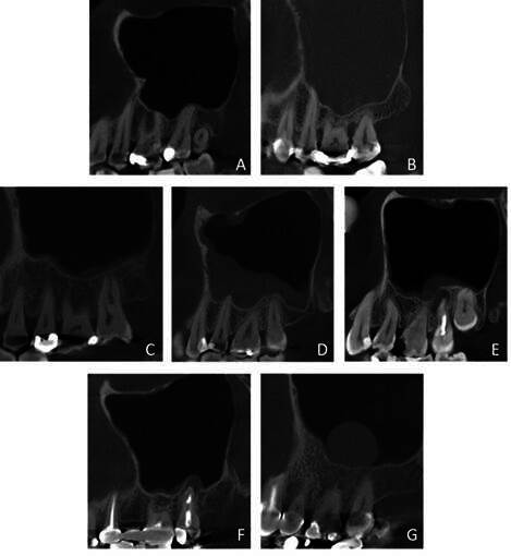
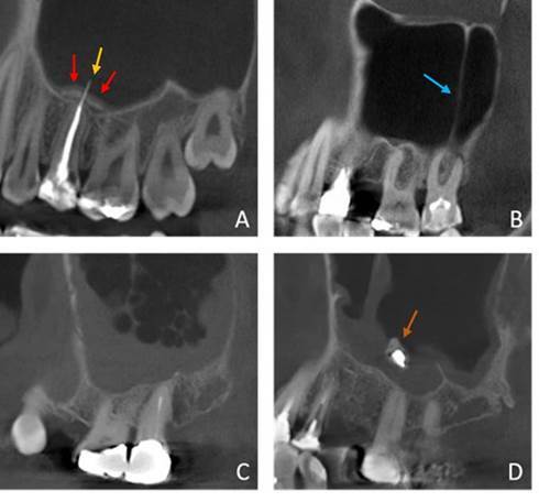
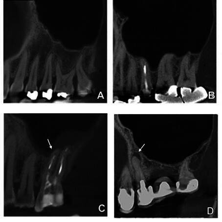
本研究旨在通过对锥形束计算机断层扫描(CBCT)图像进行回顾性分析,评估施奈德膜增厚与根尖周病理学之间的关系。为此,我们对包含 258 个窦和 1,181 颗牙齿的 147 张 CBCT 扫描图像进行了评估。硬膜层中断、牙周韧带间隙增宽、根尖牙周炎(AP)以及与 AP 相关的上颌窦底部分脱矿均被视为根尖周病变。上颌窦粘膜增厚(MSMT)分为牙源性和非牙源性。与病灶牙齿相关的不规则带状增厚和与牙根相关的局部增厚被认为是牙源性类型的 MSMT。根尖周病变的影像学特征与 MSMT 的类型和厚度之间的关系分别通过逻辑回归和线性混合模型来确定。此外,线性回归和曼-惠特尼检验评估了 AP 病变与窦底脱矿的关系(P≤0.05)。当上颌窦内存在根尖周病变时,发生牙源性 MSMT 的几率明显更高。82%的向窦底部分脱矿的 AP 与牙源性 MSMT 相关。这两种 AP 病变都部分向窦底脱矿,随着直径的增加,导致 MSMT 增加。总之,如果存在部分向窦底脱矿的 AP,则发生牙源性 MSMT 的风险为 82%。当 AP 病变较大且窦底部分脱矿时,上颌窦粘膜会增厚。
The relation between Schneiderian membrane thickening and radiodiagnostic features of periapical pathology.
This study aimed to assess the relationship between Schneiderian membrane thickening and periapical pathology in a retrospective analysis of Cone Beam Computed Tomography (CBCT) images. For this, 147 CBCT scans containing 258 sinuses and 1,181 teeth were assessed. Discontinuation of the lamina dura, widening of the periodontal ligament space, apical periodontitis (AP), and partly demineralized maxillary sinus floor associated with AP were considered periapical pathology. Maxillary sinus mucosal thickening (MSMT) was classified as odontogenic or non-odontogenic. An irregular band with a focal tooth associated thickening and local thickening related to a root were considered odontogenic types of MSMT. The relation between the imaging features of periapical pathology and the type and thickness of MSMT was determined by logistic regression and linear mixed model, respectively. In addition, linear regression and Mann Whitney test evaluated the relation and demineralization of the AP lesion towards the sinus floor (p≤0.05). The odds of having an odontogenic type of MSMT were significantly higher when a periapical pathology was present in the maxillary sinus. Eighty-two percent of AP partly demineralized towards the sinus floor were associated with an odontogenic MSMT. Both AP lesions partly demineralized towards the sinus floor and, with increased diameter, led to increased MSMT. In conclusion, there is an 82% risk of having an odontogenic type of MSMT with the presence of AP partly demineralized towards the sinus floor. More thickening of the maxillary sinus mucosa is seen with larger AP lesions and partial demineralization of the sinus floor.

 求助内容:
求助内容: 应助结果提醒方式:


