Fabiana Vitória Ananias Gonçalves, Orlando Aguirre Guedes, Sávio Akio Kachiyama, Larissa Pinzan Flauzino, Aurélio Rosa da Silva Júnior, Andreza Maria Fábio Aranha
{"title":"评估儿童牙科根管充填材料引起的牙齿变色。","authors":"Fabiana Vitória Ananias Gonçalves, Orlando Aguirre Guedes, Sávio Akio Kachiyama, Larissa Pinzan Flauzino, Aurélio Rosa da Silva Júnior, Andreza Maria Fábio Aranha","doi":"10.1590/0103-6440202405838","DOIUrl":null,"url":null,"abstract":"<p><p>This study investigated the potential for tooth discoloration of root canal filling pastes used in pediatric dentistry. Sixty bovine incisors were sectioned 2 mm apical to the cementoenamel junction and allocated into 6 groups (n = 10) according to the type of filling material used: G1- Zinc oxide-eugenol sealer; G2- Zinc oxide-eugenol and iodoform paste; G3- Calcium hydroxide (CH) and zinc oxide paste; G4- CH, zinc oxide, and iodoform paste; G5- CH and iodoform paste; and G6- Control. Polyethylene glycol 400 was used as a vehicle for CH-containing pastes. Color measurements were taken at specific intervals: preceding endodontic treatment (T0) and at successive points of 1 month (T1), 2 months (T2), 3 months (T3), and 1 year (T4) after the placement of the filling material. The color change (∆E) was calculated using the CIELab formula. Statistical analysis was performed using ANOVA, followed by Tukey's post hoc test (α = 5%). Significant differences were observed among the filling materials and time intervals (p <0.001). All groups exhibited color changes over time, except G1 and G5, which showed color changes only after 1 year. G1 and G2 demonstrated the highest ∆E values, with a statistically significant difference observed only at T2 when compared to G3 (p = 0.008). Root canal filling materials used in primary teeth have the potential to induce tooth discoloration.</p>","PeriodicalId":101363,"journal":{"name":"Brazilian dental journal","volume":"35 ","pages":"e245838"},"PeriodicalIF":0.0000,"publicationDate":"2024-09-16","publicationTypes":"Journal Article","fieldsOfStudy":null,"isOpenAccess":false,"openAccessPdf":"https://www.ncbi.nlm.nih.gov/pmc/articles/PMC11405006/pdf/","citationCount":"0","resultStr":"{\"title\":\"Assessment of tooth discoloration induced by root canal filling materials in pediatric dentistry.\",\"authors\":\"Fabiana Vitória Ananias Gonçalves, Orlando Aguirre Guedes, Sávio Akio Kachiyama, Larissa Pinzan Flauzino, Aurélio Rosa da Silva Júnior, Andreza Maria Fábio Aranha\",\"doi\":\"10.1590/0103-6440202405838\",\"DOIUrl\":null,\"url\":null,\"abstract\":\"<p><p>This study investigated the potential for tooth discoloration of root canal filling pastes used in pediatric dentistry. Sixty bovine incisors were sectioned 2 mm apical to the cementoenamel junction and allocated into 6 groups (n = 10) according to the type of filling material used: G1- Zinc oxide-eugenol sealer; G2- Zinc oxide-eugenol and iodoform paste; G3- Calcium hydroxide (CH) and zinc oxide paste; G4- CH, zinc oxide, and iodoform paste; G5- CH and iodoform paste; and G6- Control. Polyethylene glycol 400 was used as a vehicle for CH-containing pastes. Color measurements were taken at specific intervals: preceding endodontic treatment (T0) and at successive points of 1 month (T1), 2 months (T2), 3 months (T3), and 1 year (T4) after the placement of the filling material. The color change (∆E) was calculated using the CIELab formula. Statistical analysis was performed using ANOVA, followed by Tukey's post hoc test (α = 5%). Significant differences were observed among the filling materials and time intervals (p <0.001). All groups exhibited color changes over time, except G1 and G5, which showed color changes only after 1 year. G1 and G2 demonstrated the highest ∆E values, with a statistically significant difference observed only at T2 when compared to G3 (p = 0.008). Root canal filling materials used in primary teeth have the potential to induce tooth discoloration.</p>\",\"PeriodicalId\":101363,\"journal\":{\"name\":\"Brazilian dental journal\",\"volume\":\"35 \",\"pages\":\"e245838\"},\"PeriodicalIF\":0.0000,\"publicationDate\":\"2024-09-16\",\"publicationTypes\":\"Journal Article\",\"fieldsOfStudy\":null,\"isOpenAccess\":false,\"openAccessPdf\":\"https://www.ncbi.nlm.nih.gov/pmc/articles/PMC11405006/pdf/\",\"citationCount\":\"0\",\"resultStr\":null,\"platform\":\"Semanticscholar\",\"paperid\":null,\"PeriodicalName\":\"Brazilian dental journal\",\"FirstCategoryId\":\"1085\",\"ListUrlMain\":\"https://doi.org/10.1590/0103-6440202405838\",\"RegionNum\":0,\"RegionCategory\":null,\"ArticlePicture\":[],\"TitleCN\":null,\"AbstractTextCN\":null,\"PMCID\":null,\"EPubDate\":\"2024/1/1 0:00:00\",\"PubModel\":\"eCollection\",\"JCR\":\"\",\"JCRName\":\"\",\"Score\":null,\"Total\":0}","platform":"Semanticscholar","paperid":null,"PeriodicalName":"Brazilian dental journal","FirstCategoryId":"1085","ListUrlMain":"https://doi.org/10.1590/0103-6440202405838","RegionNum":0,"RegionCategory":null,"ArticlePicture":[],"TitleCN":null,"AbstractTextCN":null,"PMCID":null,"EPubDate":"2024/1/1 0:00:00","PubModel":"eCollection","JCR":"","JCRName":"","Score":null,"Total":0}
Assessment of tooth discoloration induced by root canal filling materials in pediatric dentistry.
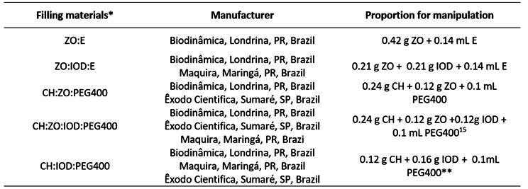
This study investigated the potential for tooth discoloration of root canal filling pastes used in pediatric dentistry. Sixty bovine incisors were sectioned 2 mm apical to the cementoenamel junction and allocated into 6 groups (n = 10) according to the type of filling material used: G1- Zinc oxide-eugenol sealer; G2- Zinc oxide-eugenol and iodoform paste; G3- Calcium hydroxide (CH) and zinc oxide paste; G4- CH, zinc oxide, and iodoform paste; G5- CH and iodoform paste; and G6- Control. Polyethylene glycol 400 was used as a vehicle for CH-containing pastes. Color measurements were taken at specific intervals: preceding endodontic treatment (T0) and at successive points of 1 month (T1), 2 months (T2), 3 months (T3), and 1 year (T4) after the placement of the filling material. The color change (∆E) was calculated using the CIELab formula. Statistical analysis was performed using ANOVA, followed by Tukey's post hoc test (α = 5%). Significant differences were observed among the filling materials and time intervals (p <0.001). All groups exhibited color changes over time, except G1 and G5, which showed color changes only after 1 year. G1 and G2 demonstrated the highest ∆E values, with a statistically significant difference observed only at T2 when compared to G3 (p = 0.008). Root canal filling materials used in primary teeth have the potential to induce tooth discoloration.

 求助内容:
求助内容: 应助结果提醒方式:


