Sarita Saraswati, Paula Martínez, Rosa Serrano, Diego Mejías, Osvaldo Graña-Castro, Ruth Álvarez Díaz, Maria A. Blasco
{"title":"肾脏成纤维细胞参与了与端粒功能障碍相关的肾脏纤维化的纤维化变化。","authors":"Sarita Saraswati, Paula Martínez, Rosa Serrano, Diego Mejías, Osvaldo Graña-Castro, Ruth Álvarez Díaz, Maria A. Blasco","doi":"10.1038/s12276-024-01318-8","DOIUrl":null,"url":null,"abstract":"Tubulointerstitial fibrosis associated with chronic kidney disease (CKD) represents a global health care problem. We previously reported that short and dysfunctional telomeres lead to interstitial renal fibrosis; however, the cell-of-origin of kidney fibrosis associated with telomere dysfunction is currently unknown. We induced telomere dysfunction by deleting the Trf1 gene encoding a telomere-binding factor specifically in renal fibroblasts in both short-term and long-term life-long experiments in mice to identify the role of fibroblasts in renal fibrosis. Short-term Trf1 deletion in renal fibroblasts was not sufficient to trigger kidney fibrosis but was sufficient to induce inflammatory responses, ECM deposition, cell cycle arrest, fibrogenesis, and vascular rarefaction. However, long-term persistent deletion of Trf1 in fibroblasts resulted in kidney fibrosis accompanied by an elevated urinary albumin-to-creatinine ratio (uACR) and a decrease in mouse survival. These cellular responses lead to the macrophage-to-myofibroblast transition (MMT), endothelial-to-mesenchymal transition (EndMT), and partial epithelial-to-mesenchymal transition (EMT), ultimately causing kidney fibrosis at the humane endpoint (HEP) when the deletion of Trf1 in fibroblasts is maintained throughout the lifespan of mice. Our findings contribute to a better understanding of the role of dysfunctional telomeres in the onset of the profibrotic alterations that lead to kidney fibrosis. Chronic kidney disease, a condition that can lead to kidney failure and death, affects millions worldwide. It’s characterized by kidney fibrosis, a process where healthy kidney tissue becomes scar tissue. The exact cells that start this process were unknown. Researchers studied the role of a protein called TRF1, which protects the ends of chromosomes, in kidney fibroblasts, cells that make connective tissue. They removed TRF1 from these cells in mice and observed the effects. Results showed that removing TRF1 from fibroblasts increased fibrosis, inflammation, and kidney damage. Specifically, fibroblasts without TRF1 were more likely to change into myofibroblasts, leading to more scar tissue. Study concluded that TRF1 is vital in preventing kidney fibrosis by keeping fibroblasts healthy. This finding improves our understanding of CKD and suggests potential treatments targeting fibroblasts and TRF1 to fight kidney fibrosis. This summary was initially drafted using artificial intelligence, then revised and fact-checked by the author.","PeriodicalId":50466,"journal":{"name":"Experimental and Molecular Medicine","volume":null,"pages":null},"PeriodicalIF":9.5000,"publicationDate":"2024-10-01","publicationTypes":"Journal Article","fieldsOfStudy":null,"isOpenAccess":false,"openAccessPdf":"https://www.nature.com/articles/s12276-024-01318-8.pdf","citationCount":"0","resultStr":"{\"title\":\"Renal fibroblasts are involved in fibrogenic changes in kidney fibrosis associated with dysfunctional telomeres\",\"authors\":\"Sarita Saraswati, Paula Martínez, Rosa Serrano, Diego Mejías, Osvaldo Graña-Castro, Ruth Álvarez Díaz, Maria A. Blasco\",\"doi\":\"10.1038/s12276-024-01318-8\",\"DOIUrl\":null,\"url\":null,\"abstract\":\"Tubulointerstitial fibrosis associated with chronic kidney disease (CKD) represents a global health care problem. We previously reported that short and dysfunctional telomeres lead to interstitial renal fibrosis; however, the cell-of-origin of kidney fibrosis associated with telomere dysfunction is currently unknown. We induced telomere dysfunction by deleting the Trf1 gene encoding a telomere-binding factor specifically in renal fibroblasts in both short-term and long-term life-long experiments in mice to identify the role of fibroblasts in renal fibrosis. Short-term Trf1 deletion in renal fibroblasts was not sufficient to trigger kidney fibrosis but was sufficient to induce inflammatory responses, ECM deposition, cell cycle arrest, fibrogenesis, and vascular rarefaction. However, long-term persistent deletion of Trf1 in fibroblasts resulted in kidney fibrosis accompanied by an elevated urinary albumin-to-creatinine ratio (uACR) and a decrease in mouse survival. These cellular responses lead to the macrophage-to-myofibroblast transition (MMT), endothelial-to-mesenchymal transition (EndMT), and partial epithelial-to-mesenchymal transition (EMT), ultimately causing kidney fibrosis at the humane endpoint (HEP) when the deletion of Trf1 in fibroblasts is maintained throughout the lifespan of mice. Our findings contribute to a better understanding of the role of dysfunctional telomeres in the onset of the profibrotic alterations that lead to kidney fibrosis. Chronic kidney disease, a condition that can lead to kidney failure and death, affects millions worldwide. It’s characterized by kidney fibrosis, a process where healthy kidney tissue becomes scar tissue. The exact cells that start this process were unknown. Researchers studied the role of a protein called TRF1, which protects the ends of chromosomes, in kidney fibroblasts, cells that make connective tissue. They removed TRF1 from these cells in mice and observed the effects. Results showed that removing TRF1 from fibroblasts increased fibrosis, inflammation, and kidney damage. Specifically, fibroblasts without TRF1 were more likely to change into myofibroblasts, leading to more scar tissue. Study concluded that TRF1 is vital in preventing kidney fibrosis by keeping fibroblasts healthy. This finding improves our understanding of CKD and suggests potential treatments targeting fibroblasts and TRF1 to fight kidney fibrosis. This summary was initially drafted using artificial intelligence, then revised and fact-checked by the author.\",\"PeriodicalId\":50466,\"journal\":{\"name\":\"Experimental and Molecular Medicine\",\"volume\":null,\"pages\":null},\"PeriodicalIF\":9.5000,\"publicationDate\":\"2024-10-01\",\"publicationTypes\":\"Journal Article\",\"fieldsOfStudy\":null,\"isOpenAccess\":false,\"openAccessPdf\":\"https://www.nature.com/articles/s12276-024-01318-8.pdf\",\"citationCount\":\"0\",\"resultStr\":null,\"platform\":\"Semanticscholar\",\"paperid\":null,\"PeriodicalName\":\"Experimental and Molecular Medicine\",\"FirstCategoryId\":\"3\",\"ListUrlMain\":\"https://www.nature.com/articles/s12276-024-01318-8\",\"RegionNum\":2,\"RegionCategory\":\"医学\",\"ArticlePicture\":[],\"TitleCN\":null,\"AbstractTextCN\":null,\"PMCID\":null,\"EPubDate\":\"\",\"PubModel\":\"\",\"JCR\":\"Q1\",\"JCRName\":\"BIOCHEMISTRY & MOLECULAR BIOLOGY\",\"Score\":null,\"Total\":0}","platform":"Semanticscholar","paperid":null,"PeriodicalName":"Experimental and Molecular Medicine","FirstCategoryId":"3","ListUrlMain":"https://www.nature.com/articles/s12276-024-01318-8","RegionNum":2,"RegionCategory":"医学","ArticlePicture":[],"TitleCN":null,"AbstractTextCN":null,"PMCID":null,"EPubDate":"","PubModel":"","JCR":"Q1","JCRName":"BIOCHEMISTRY & MOLECULAR BIOLOGY","Score":null,"Total":0}
Renal fibroblasts are involved in fibrogenic changes in kidney fibrosis associated with dysfunctional telomeres
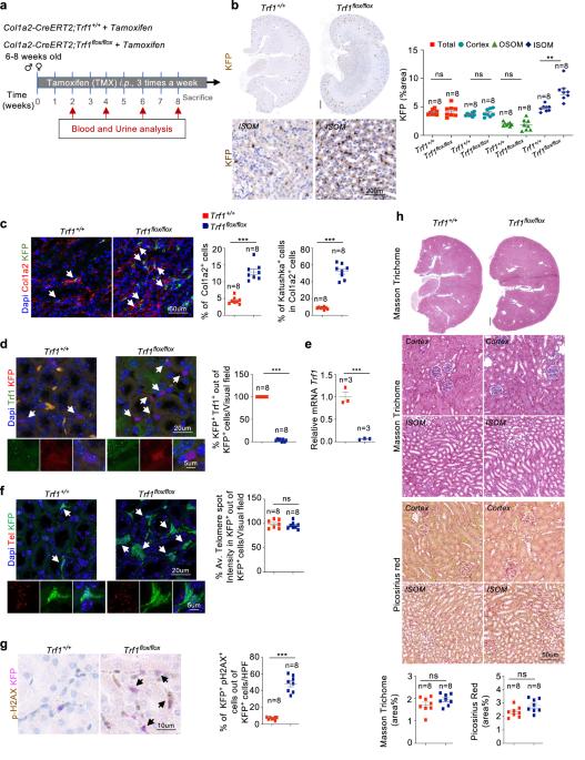
Tubulointerstitial fibrosis associated with chronic kidney disease (CKD) represents a global health care problem. We previously reported that short and dysfunctional telomeres lead to interstitial renal fibrosis; however, the cell-of-origin of kidney fibrosis associated with telomere dysfunction is currently unknown. We induced telomere dysfunction by deleting the Trf1 gene encoding a telomere-binding factor specifically in renal fibroblasts in both short-term and long-term life-long experiments in mice to identify the role of fibroblasts in renal fibrosis. Short-term Trf1 deletion in renal fibroblasts was not sufficient to trigger kidney fibrosis but was sufficient to induce inflammatory responses, ECM deposition, cell cycle arrest, fibrogenesis, and vascular rarefaction. However, long-term persistent deletion of Trf1 in fibroblasts resulted in kidney fibrosis accompanied by an elevated urinary albumin-to-creatinine ratio (uACR) and a decrease in mouse survival. These cellular responses lead to the macrophage-to-myofibroblast transition (MMT), endothelial-to-mesenchymal transition (EndMT), and partial epithelial-to-mesenchymal transition (EMT), ultimately causing kidney fibrosis at the humane endpoint (HEP) when the deletion of Trf1 in fibroblasts is maintained throughout the lifespan of mice. Our findings contribute to a better understanding of the role of dysfunctional telomeres in the onset of the profibrotic alterations that lead to kidney fibrosis. Chronic kidney disease, a condition that can lead to kidney failure and death, affects millions worldwide. It’s characterized by kidney fibrosis, a process where healthy kidney tissue becomes scar tissue. The exact cells that start this process were unknown. Researchers studied the role of a protein called TRF1, which protects the ends of chromosomes, in kidney fibroblasts, cells that make connective tissue. They removed TRF1 from these cells in mice and observed the effects. Results showed that removing TRF1 from fibroblasts increased fibrosis, inflammation, and kidney damage. Specifically, fibroblasts without TRF1 were more likely to change into myofibroblasts, leading to more scar tissue. Study concluded that TRF1 is vital in preventing kidney fibrosis by keeping fibroblasts healthy. This finding improves our understanding of CKD and suggests potential treatments targeting fibroblasts and TRF1 to fight kidney fibrosis. This summary was initially drafted using artificial intelligence, then revised and fact-checked by the author.
期刊介绍:
Experimental & Molecular Medicine (EMM) stands as Korea's pioneering biochemistry journal, established in 1964 and rejuvenated in 1996 as an Open Access, fully peer-reviewed international journal. Dedicated to advancing translational research and showcasing recent breakthroughs in the biomedical realm, EMM invites submissions encompassing genetic, molecular, and cellular studies of human physiology and diseases. Emphasizing the correlation between experimental and translational research and enhanced clinical benefits, the journal actively encourages contributions employing specific molecular tools. Welcoming studies that bridge basic discoveries with clinical relevance, alongside articles demonstrating clear in vivo significance and novelty, Experimental & Molecular Medicine proudly serves as an open-access, online-only repository of cutting-edge medical research.