Aynur Azizova, Ivar J. H. G. Wamelink, Yeva Prysiazhniuk, Marcus Cakmak, Elif Kaya, Jan Petr, Frederik Barkhof, Vera C. Keil
{"title":"利用无钆磁共振成像序列预测胶质瘤增强质量的人类表现。","authors":"Aynur Azizova, Ivar J. H. G. Wamelink, Yeva Prysiazhniuk, Marcus Cakmak, Elif Kaya, Jan Petr, Frederik Barkhof, Vera C. Keil","doi":"10.1111/jon.13233","DOIUrl":null,"url":null,"abstract":"<div>\n \n \n <section>\n \n <h3> Background and Purpose</h3>\n \n <p>To develop and test a decision tree for predicting contrast enhancement quality and shape using precontrast magnetic resonance imaging (MRI) sequences in a large adult-type diffuse glioma cohort.</p>\n </section>\n \n <section>\n \n <h3> Methods</h3>\n \n <p>Preoperative MRI scans (development/optimization/test sets: <i>n</i> = 31/38/303, male = 17/22/189, mean age = 52/59/56.7 years, high-grade glioma = 22/33/249) were retrospectively evaluated, including pre- and postcontrast T1-weighted, T2-weighted, fluid-attenuated inversion recovery, and diffusion-weighted imaging sequences. Enhancement prediction decision tree (EPDT) was developed using development and optimization sets, incorporating four imaging features: necrosis, diffusion restriction, T2 inhomogeneity, and nonenhancing tumor margins. EPDT accuracy was assessed on a test set by three raters of variable experience. True enhancement features (gold standard) were evaluated using pre- and postcontrast T1-weighted images. Statistical analysis used confusion matrices, Cohen's/Fleiss’ kappa, and Kendall's <i>W</i>. Significance threshold was <i>p</i> < .05.</p>\n </section>\n \n <section>\n \n <h3> Results</h3>\n \n <p>Raters 1, 2, and 3 achieved overall accuracies of .86 (95% confidence interval [CI]: .81-.90), .89 (95% CI: .85-.92), and .92 (95% CI: .89-.95), respectively, in predicting enhancement quality (marked, mild, or no enhancement). Regarding shape, defined as the thickness of enhancing margin (solid, rim, or no enhancement), accuracies were .84 (95% CI: .79-.88), .88 (95% CI: .84-.92), and .89 (95% CI: .85-.92). Intrarater intergroup agreement comparing predicted and true enhancement features consistently reached substantial levels (≥.68 [95% CI: .61-.75]). Interrater comparison showed at least moderate agreement (group: ≥.42 [95% CI: .36-.48], pairwise: ≥.61 [95% CI: .50-.72]). Among the imaging features in the EPDT, necrosis assessment displayed the highest intra- and interrater consistency (≥.80 [95% CI: .73-.88]).</p>\n </section>\n \n <section>\n \n <h3> Conclusion</h3>\n \n <p>The proposed EPDT has high accuracy in predicting enhancement patterns of gliomas irrespective of rater experience.</p>\n </section>\n </div>","PeriodicalId":16399,"journal":{"name":"Journal of Neuroimaging","volume":"34 6","pages":"673-693"},"PeriodicalIF":2.3000,"publicationDate":"2024-09-19","publicationTypes":"Journal Article","fieldsOfStudy":null,"isOpenAccess":false,"openAccessPdf":"https://onlinelibrary.wiley.com/doi/epdf/10.1111/jon.13233","citationCount":"0","resultStr":"{\"title\":\"Human performance in predicting enhancement quality of gliomas using gadolinium-free MRI sequences\",\"authors\":\"Aynur Azizova, Ivar J. H. G. Wamelink, Yeva Prysiazhniuk, Marcus Cakmak, Elif Kaya, Jan Petr, Frederik Barkhof, Vera C. Keil\",\"doi\":\"10.1111/jon.13233\",\"DOIUrl\":null,\"url\":null,\"abstract\":\"<div>\\n \\n \\n <section>\\n \\n <h3> Background and Purpose</h3>\\n \\n <p>To develop and test a decision tree for predicting contrast enhancement quality and shape using precontrast magnetic resonance imaging (MRI) sequences in a large adult-type diffuse glioma cohort.</p>\\n </section>\\n \\n <section>\\n \\n <h3> Methods</h3>\\n \\n <p>Preoperative MRI scans (development/optimization/test sets: <i>n</i> = 31/38/303, male = 17/22/189, mean age = 52/59/56.7 years, high-grade glioma = 22/33/249) were retrospectively evaluated, including pre- and postcontrast T1-weighted, T2-weighted, fluid-attenuated inversion recovery, and diffusion-weighted imaging sequences. Enhancement prediction decision tree (EPDT) was developed using development and optimization sets, incorporating four imaging features: necrosis, diffusion restriction, T2 inhomogeneity, and nonenhancing tumor margins. EPDT accuracy was assessed on a test set by three raters of variable experience. True enhancement features (gold standard) were evaluated using pre- and postcontrast T1-weighted images. Statistical analysis used confusion matrices, Cohen's/Fleiss’ kappa, and Kendall's <i>W</i>. Significance threshold was <i>p</i> < .05.</p>\\n </section>\\n \\n <section>\\n \\n <h3> Results</h3>\\n \\n <p>Raters 1, 2, and 3 achieved overall accuracies of .86 (95% confidence interval [CI]: .81-.90), .89 (95% CI: .85-.92), and .92 (95% CI: .89-.95), respectively, in predicting enhancement quality (marked, mild, or no enhancement). Regarding shape, defined as the thickness of enhancing margin (solid, rim, or no enhancement), accuracies were .84 (95% CI: .79-.88), .88 (95% CI: .84-.92), and .89 (95% CI: .85-.92). Intrarater intergroup agreement comparing predicted and true enhancement features consistently reached substantial levels (≥.68 [95% CI: .61-.75]). Interrater comparison showed at least moderate agreement (group: ≥.42 [95% CI: .36-.48], pairwise: ≥.61 [95% CI: .50-.72]). Among the imaging features in the EPDT, necrosis assessment displayed the highest intra- and interrater consistency (≥.80 [95% CI: .73-.88]).</p>\\n </section>\\n \\n <section>\\n \\n <h3> Conclusion</h3>\\n \\n <p>The proposed EPDT has high accuracy in predicting enhancement patterns of gliomas irrespective of rater experience.</p>\\n </section>\\n </div>\",\"PeriodicalId\":16399,\"journal\":{\"name\":\"Journal of Neuroimaging\",\"volume\":\"34 6\",\"pages\":\"673-693\"},\"PeriodicalIF\":2.3000,\"publicationDate\":\"2024-09-19\",\"publicationTypes\":\"Journal Article\",\"fieldsOfStudy\":null,\"isOpenAccess\":false,\"openAccessPdf\":\"https://onlinelibrary.wiley.com/doi/epdf/10.1111/jon.13233\",\"citationCount\":\"0\",\"resultStr\":null,\"platform\":\"Semanticscholar\",\"paperid\":null,\"PeriodicalName\":\"Journal of Neuroimaging\",\"FirstCategoryId\":\"3\",\"ListUrlMain\":\"https://onlinelibrary.wiley.com/doi/10.1111/jon.13233\",\"RegionNum\":4,\"RegionCategory\":\"医学\",\"ArticlePicture\":[],\"TitleCN\":null,\"AbstractTextCN\":null,\"PMCID\":null,\"EPubDate\":\"\",\"PubModel\":\"\",\"JCR\":\"Q3\",\"JCRName\":\"CLINICAL NEUROLOGY\",\"Score\":null,\"Total\":0}","platform":"Semanticscholar","paperid":null,"PeriodicalName":"Journal of Neuroimaging","FirstCategoryId":"3","ListUrlMain":"https://onlinelibrary.wiley.com/doi/10.1111/jon.13233","RegionNum":4,"RegionCategory":"医学","ArticlePicture":[],"TitleCN":null,"AbstractTextCN":null,"PMCID":null,"EPubDate":"","PubModel":"","JCR":"Q3","JCRName":"CLINICAL NEUROLOGY","Score":null,"Total":0}
Human performance in predicting enhancement quality of gliomas using gadolinium-free MRI sequences
Background and Purpose
To develop and test a decision tree for predicting contrast enhancement quality and shape using precontrast magnetic resonance imaging (MRI) sequences in a large adult-type diffuse glioma cohort.
Methods
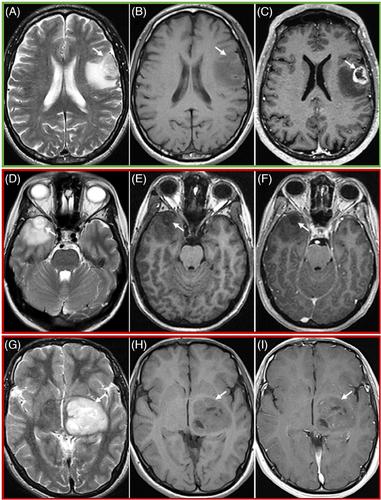
Preoperative MRI scans (development/optimization/test sets: n = 31/38/303, male = 17/22/189, mean age = 52/59/56.7 years, high-grade glioma = 22/33/249) were retrospectively evaluated, including pre- and postcontrast T1-weighted, T2-weighted, fluid-attenuated inversion recovery, and diffusion-weighted imaging sequences. Enhancement prediction decision tree (EPDT) was developed using development and optimization sets, incorporating four imaging features: necrosis, diffusion restriction, T2 inhomogeneity, and nonenhancing tumor margins. EPDT accuracy was assessed on a test set by three raters of variable experience. True enhancement features (gold standard) were evaluated using pre- and postcontrast T1-weighted images. Statistical analysis used confusion matrices, Cohen's/Fleiss’ kappa, and Kendall's W. Significance threshold was p < .05.
Results
Raters 1, 2, and 3 achieved overall accuracies of .86 (95% confidence interval [CI]: .81-.90), .89 (95% CI: .85-.92), and .92 (95% CI: .89-.95), respectively, in predicting enhancement quality (marked, mild, or no enhancement). Regarding shape, defined as the thickness of enhancing margin (solid, rim, or no enhancement), accuracies were .84 (95% CI: .79-.88), .88 (95% CI: .84-.92), and .89 (95% CI: .85-.92). Intrarater intergroup agreement comparing predicted and true enhancement features consistently reached substantial levels (≥.68 [95% CI: .61-.75]). Interrater comparison showed at least moderate agreement (group: ≥.42 [95% CI: .36-.48], pairwise: ≥.61 [95% CI: .50-.72]). Among the imaging features in the EPDT, necrosis assessment displayed the highest intra- and interrater consistency (≥.80 [95% CI: .73-.88]).
Conclusion
The proposed EPDT has high accuracy in predicting enhancement patterns of gliomas irrespective of rater experience.
期刊介绍:
Start reading the Journal of Neuroimaging to learn the latest neurological imaging techniques. The peer-reviewed research is written in a practical clinical context, giving you the information you need on:
MRI
CT
Carotid Ultrasound and TCD
SPECT
PET
Endovascular Surgical Neuroradiology
Functional MRI
Xenon CT
and other new and upcoming neuroscientific modalities.The Journal of Neuroimaging addresses the full spectrum of human nervous system disease, including stroke, neoplasia, degenerating and demyelinating disease, epilepsy, tumors, lesions, infectious disease, cerebral vascular arterial diseases, toxic-metabolic disease, psychoses, dementias, heredo-familial disease, and trauma.Offering original research, review articles, case reports, neuroimaging CPCs, and evaluations of instruments and technology relevant to the nervous system, the Journal of Neuroimaging focuses on useful clinical developments and applications, tested techniques and interpretations, patient care, diagnostics, and therapeutics. Start reading today!

 求助内容:
求助内容: 应助结果提醒方式:


