Severin K. Lustenberger , Claudia A. Castro Jaramillo , Lena A. Bärtschi , Rich Williams , Roger Schibli , Linjing Mu , Stefanie D. Krämer
{"title":"通过 PET 对癌症免疫状态进行成像:用携带氰基前端的 11C 标记 P1-Asn 拟肽物靶向豆豆蛋白酶","authors":"Severin K. Lustenberger , Claudia A. Castro Jaramillo , Lena A. Bärtschi , Rich Williams , Roger Schibli , Linjing Mu , Stefanie D. Krämer","doi":"10.1016/j.nucmedbio.2024.108951","DOIUrl":null,"url":null,"abstract":"<div><h3>Purpose</h3><p>M2-type tumor-associated macrophages (TAM) residing in the tumor microenvironment (TME) have been linked to tumor invasiveness, metastasis and poor prognosis. M2 TAMs suppress T cell activation, silencing the recognition of the cancer by the immune system. Targeting TAMs in anti-cancer therapy may support the immune system and immune-checkpoint inhibitor therapies to fight the cancer cells. We aimed to develop a PET tracer for the imaging of M2 TAM infiltration of cancer, using activated legumain as the imaging target.</p></div><div><h3>Basic procedures</h3><p>Two P1-mimicking inhibitors with a cyano-warhead were labeled with carbon-11 and evaluated <em>in vitro</em> and <em>in vivo</em> with a CT26 tumor mouse model. Target expression and activity were quantified from RT-qPCR and <em>in vitro</em> substrate conversion, respectively. The co-localization of legumain and TAMs was assessed by fluorescence microscopy. The two tracers were evaluated by PET with subsequent biodistribution analysis with the dissected tissues. Parent-to-total radioactivity in plasma was determined at several time points after i.v. tracer injection, using reverse phase radio-UPLC.</p></div><div><h3>Main findings</h3><p>Legumain displayed a target density of 40.7 ± 19.1 pmol per mg total protein in tumor lysate (<em>n</em> = 4) with high substrate conversion and colocalization with M2 macrophages in the tumor periphery. [<sup>11</sup>C]<strong>1</strong> and [<sup>11</sup>C]<strong>2</strong> were synthesized with >95 % radiochemical purity and 12.9–382.2 GBq/μmol molar activity at the end of synthesis. We observed heterogeneous tumor accumulation in <em>in vitro</em> autoradiography and PET for both tracers. However, excess unlabeled <strong>1</strong> or <strong>2</strong> did not compete with tracer accumulation. Both [<sup>11</sup>C]<strong>1</strong> and [<sup>11</sup>C]<strong>2</strong> were rapidly metabolized to a polar radiometabolite <em>in vivo</em>.</p></div><div><h3>Principal conclusions</h3><p>The legumain tracers [<sup>11</sup>C]<strong>1</strong> and [<sup>11</sup>C]<strong>2</strong>, synthesized with high radiochemical purity and molar activity, accumulate in the legumain-positive CT26 tumor <em>in vivo</em>. However, the lack of competition by excess compound questions their specificity. Both tracers are rapidly metabolized <em>in vivo</em>, requiring structural modifications towards more stable tracers for further investigations.</p></div>","PeriodicalId":19363,"journal":{"name":"Nuclear medicine and biology","volume":"138 ","pages":"Article 108951"},"PeriodicalIF":3.6000,"publicationDate":"2024-09-13","publicationTypes":"Journal Article","fieldsOfStudy":null,"isOpenAccess":false,"openAccessPdf":"https://www.sciencedirect.com/science/article/pii/S0969805124000775/pdfft?md5=e565835330dda2cc416d9d587895cf69&pid=1-s2.0-S0969805124000775-main.pdf","citationCount":"0","resultStr":"{\"title\":\"Towards imaging the immune state of cancer by PET: Targeting legumain with 11C-labeled P1-Asn peptidomimetics carrying a cyano-warhead\",\"authors\":\"Severin K. Lustenberger , Claudia A. Castro Jaramillo , Lena A. Bärtschi , Rich Williams , Roger Schibli , Linjing Mu , Stefanie D. Krämer\",\"doi\":\"10.1016/j.nucmedbio.2024.108951\",\"DOIUrl\":null,\"url\":null,\"abstract\":\"<div><h3>Purpose</h3><p>M2-type tumor-associated macrophages (TAM) residing in the tumor microenvironment (TME) have been linked to tumor invasiveness, metastasis and poor prognosis. M2 TAMs suppress T cell activation, silencing the recognition of the cancer by the immune system. Targeting TAMs in anti-cancer therapy may support the immune system and immune-checkpoint inhibitor therapies to fight the cancer cells. We aimed to develop a PET tracer for the imaging of M2 TAM infiltration of cancer, using activated legumain as the imaging target.</p></div><div><h3>Basic procedures</h3><p>Two P1-mimicking inhibitors with a cyano-warhead were labeled with carbon-11 and evaluated <em>in vitro</em> and <em>in vivo</em> with a CT26 tumor mouse model. Target expression and activity were quantified from RT-qPCR and <em>in vitro</em> substrate conversion, respectively. The co-localization of legumain and TAMs was assessed by fluorescence microscopy. The two tracers were evaluated by PET with subsequent biodistribution analysis with the dissected tissues. Parent-to-total radioactivity in plasma was determined at several time points after i.v. tracer injection, using reverse phase radio-UPLC.</p></div><div><h3>Main findings</h3><p>Legumain displayed a target density of 40.7 ± 19.1 pmol per mg total protein in tumor lysate (<em>n</em> = 4) with high substrate conversion and colocalization with M2 macrophages in the tumor periphery. [<sup>11</sup>C]<strong>1</strong> and [<sup>11</sup>C]<strong>2</strong> were synthesized with >95 % radiochemical purity and 12.9–382.2 GBq/μmol molar activity at the end of synthesis. We observed heterogeneous tumor accumulation in <em>in vitro</em> autoradiography and PET for both tracers. However, excess unlabeled <strong>1</strong> or <strong>2</strong> did not compete with tracer accumulation. Both [<sup>11</sup>C]<strong>1</strong> and [<sup>11</sup>C]<strong>2</strong> were rapidly metabolized to a polar radiometabolite <em>in vivo</em>.</p></div><div><h3>Principal conclusions</h3><p>The legumain tracers [<sup>11</sup>C]<strong>1</strong> and [<sup>11</sup>C]<strong>2</strong>, synthesized with high radiochemical purity and molar activity, accumulate in the legumain-positive CT26 tumor <em>in vivo</em>. However, the lack of competition by excess compound questions their specificity. Both tracers are rapidly metabolized <em>in vivo</em>, requiring structural modifications towards more stable tracers for further investigations.</p></div>\",\"PeriodicalId\":19363,\"journal\":{\"name\":\"Nuclear medicine and biology\",\"volume\":\"138 \",\"pages\":\"Article 108951\"},\"PeriodicalIF\":3.6000,\"publicationDate\":\"2024-09-13\",\"publicationTypes\":\"Journal Article\",\"fieldsOfStudy\":null,\"isOpenAccess\":false,\"openAccessPdf\":\"https://www.sciencedirect.com/science/article/pii/S0969805124000775/pdfft?md5=e565835330dda2cc416d9d587895cf69&pid=1-s2.0-S0969805124000775-main.pdf\",\"citationCount\":\"0\",\"resultStr\":null,\"platform\":\"Semanticscholar\",\"paperid\":null,\"PeriodicalName\":\"Nuclear medicine and biology\",\"FirstCategoryId\":\"3\",\"ListUrlMain\":\"https://www.sciencedirect.com/science/article/pii/S0969805124000775\",\"RegionNum\":4,\"RegionCategory\":\"医学\",\"ArticlePicture\":[],\"TitleCN\":null,\"AbstractTextCN\":null,\"PMCID\":null,\"EPubDate\":\"\",\"PubModel\":\"\",\"JCR\":\"Q1\",\"JCRName\":\"RADIOLOGY, NUCLEAR MEDICINE & MEDICAL IMAGING\",\"Score\":null,\"Total\":0}","platform":"Semanticscholar","paperid":null,"PeriodicalName":"Nuclear medicine and biology","FirstCategoryId":"3","ListUrlMain":"https://www.sciencedirect.com/science/article/pii/S0969805124000775","RegionNum":4,"RegionCategory":"医学","ArticlePicture":[],"TitleCN":null,"AbstractTextCN":null,"PMCID":null,"EPubDate":"","PubModel":"","JCR":"Q1","JCRName":"RADIOLOGY, NUCLEAR MEDICINE & MEDICAL IMAGING","Score":null,"Total":0}
引用次数: 0
摘要
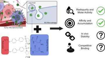
驻留在肿瘤微环境(TME)中的 M2 型肿瘤相关巨噬细胞(TAM)与肿瘤的侵袭性、转移和不良预后有关。M2 TAMs 可抑制 T 细胞的活化,从而抑制免疫系统对癌症的识别。在抗癌治疗中靶向TAMs可支持免疫系统和免疫检查点抑制剂疗法来对抗癌细胞。我们的目标是开发一种 PET 示踪剂,以活化的豆豆蛋白酶为成像靶点,对癌症中 M2 TAM 的浸润进行成像。
Towards imaging the immune state of cancer by PET: Targeting legumain with 11C-labeled P1-Asn peptidomimetics carrying a cyano-warhead
Purpose
M2-type tumor-associated macrophages (TAM) residing in the tumor microenvironment (TME) have been linked to tumor invasiveness, metastasis and poor prognosis. M2 TAMs suppress T cell activation, silencing the recognition of the cancer by the immune system. Targeting TAMs in anti-cancer therapy may support the immune system and immune-checkpoint inhibitor therapies to fight the cancer cells. We aimed to develop a PET tracer for the imaging of M2 TAM infiltration of cancer, using activated legumain as the imaging target.
Basic procedures
Two P1-mimicking inhibitors with a cyano-warhead were labeled with carbon-11 and evaluated in vitro and in vivo with a CT26 tumor mouse model. Target expression and activity were quantified from RT-qPCR and in vitro substrate conversion, respectively. The co-localization of legumain and TAMs was assessed by fluorescence microscopy. The two tracers were evaluated by PET with subsequent biodistribution analysis with the dissected tissues. Parent-to-total radioactivity in plasma was determined at several time points after i.v. tracer injection, using reverse phase radio-UPLC.
Main findings
Legumain displayed a target density of 40.7 ± 19.1 pmol per mg total protein in tumor lysate (n = 4) with high substrate conversion and colocalization with M2 macrophages in the tumor periphery. [11C]1 and [11C]2 were synthesized with >95 % radiochemical purity and 12.9–382.2 GBq/μmol molar activity at the end of synthesis. We observed heterogeneous tumor accumulation in in vitro autoradiography and PET for both tracers. However, excess unlabeled 1 or 2 did not compete with tracer accumulation. Both [11C]1 and [11C]2 were rapidly metabolized to a polar radiometabolite in vivo.
Principal conclusions
The legumain tracers [11C]1 and [11C]2, synthesized with high radiochemical purity and molar activity, accumulate in the legumain-positive CT26 tumor in vivo. However, the lack of competition by excess compound questions their specificity. Both tracers are rapidly metabolized in vivo, requiring structural modifications towards more stable tracers for further investigations.
期刊介绍:
Nuclear Medicine and Biology publishes original research addressing all aspects of radiopharmaceutical science: synthesis, in vitro and ex vivo studies, in vivo biodistribution by dissection or imaging, radiopharmacology, radiopharmacy, and translational clinical studies of new targeted radiotracers. The importance of the target to an unmet clinical need should be the first consideration. If the synthesis of a new radiopharmaceutical is submitted without in vitro or in vivo data, then the uniqueness of the chemistry must be emphasized.
These multidisciplinary studies should validate the mechanism of localization whether the probe is based on binding to a receptor, enzyme, tumor antigen, or another well-defined target. The studies should be aimed at evaluating how the chemical and radiopharmaceutical properties affect pharmacokinetics, pharmacodynamics, or therapeutic efficacy. Ideally, the study would address the sensitivity of the probe to changes in disease or treatment, although studies validating mechanism alone are acceptable. Radiopharmacy practice, addressing the issues of preparation, automation, quality control, dispensing, and regulations applicable to qualification and administration of radiopharmaceuticals to humans, is an important aspect of the developmental process, but only if the study has a significant impact on the field.
Contributions on the subject of therapeutic radiopharmaceuticals also are appropriate provided that the specificity of labeled compound localization and therapeutic effect have been addressed.
 求助内容:
求助内容: 应助结果提醒方式:


