Ragavendiran Anandan, Krithika C. Lakshmi, Anuradha Ganesan, Yesoda AniyanK
{"title":"用磁共振成像评估口腔黏膜下纤维化的咽部气道空间:横断面观察研究","authors":"Ragavendiran Anandan, Krithika C. Lakshmi, Anuradha Ganesan, Yesoda AniyanK","doi":"10.1016/j.jobcr.2024.09.003","DOIUrl":null,"url":null,"abstract":"<div><h3>Introduction</h3><p>Oral submucous fibrosis (OSMF) alters the pharynx, which may affect airway size. MRI will be useful for diagnosing pharyngeal abnormalities. MRI is used to evaluate pharyngeal airway and soft palate changes in OSMF patients.</p></div><div><h3>Materials and methods</h3><p>This study is a cross-sectional observational study that included a sample size of 42 patients. Group A consisted of 21 patients with OSMF, while Group B consisted of 21 volunteers without OSMF, who served as the control group. The patients with OSMF were classified into Stages I, II, and III according to the categorization established by Pindborg JJ in 1989, Stop-Bang questionnaire was employed to assess obstructive sleep apnoea. Magnetic Resonance Imaging (MRI) was utilized to acquire evaluations of the pharyngeal airway, encompassing measurements in the midsagittal, cross-sectional width, length planes, and cross-sectional area with volume, for all participants. The Shapiro-Wilk test determines distribution normality. We utilized one-way ANOVA to compare the means between groups.</p></div><div><h3>Results</h3><p>The average age of OSMF patients was 45.9 ± 8.16, while the control group was 39.19 ± 4.21. Stage I of OSMF had the highest mean Stop Bang questionnaire score (2.75), followed by stage III (2.22), and stage II (1.75). Statistically significant differences (p < 0.001) were seen in volume, linear midsagittal planes, cross-sectional width and length planes, cross-sectional area, and soft palate breadth and length between OSMF and control groups.</p></div><div><h3>Conclusion</h3><p>MRI can effectively examine early changes in the pharyngeal airway of patients with OSMF thereby serving as a constructive diagnostic and motivational tool.</p></div>","PeriodicalId":16609,"journal":{"name":"Journal of oral biology and craniofacial research","volume":"14 6","pages":"Pages 669-675"},"PeriodicalIF":0.0000,"publicationDate":"2024-09-19","publicationTypes":"Journal Article","fieldsOfStudy":null,"isOpenAccess":false,"openAccessPdf":"https://www.sciencedirect.com/science/article/pii/S2212426824001271/pdfft?md5=7977b3196c39aa36badc0eb3329f1a70&pid=1-s2.0-S2212426824001271-main.pdf","citationCount":"0","resultStr":"{\"title\":\"Assessment of pharyngeal airway space with MRI In oral submucous fibrosis: A cross-sectional observational study\",\"authors\":\"Ragavendiran Anandan, Krithika C. Lakshmi, Anuradha Ganesan, Yesoda AniyanK\",\"doi\":\"10.1016/j.jobcr.2024.09.003\",\"DOIUrl\":null,\"url\":null,\"abstract\":\"<div><h3>Introduction</h3><p>Oral submucous fibrosis (OSMF) alters the pharynx, which may affect airway size. MRI will be useful for diagnosing pharyngeal abnormalities. MRI is used to evaluate pharyngeal airway and soft palate changes in OSMF patients.</p></div><div><h3>Materials and methods</h3><p>This study is a cross-sectional observational study that included a sample size of 42 patients. Group A consisted of 21 patients with OSMF, while Group B consisted of 21 volunteers without OSMF, who served as the control group. The patients with OSMF were classified into Stages I, II, and III according to the categorization established by Pindborg JJ in 1989, Stop-Bang questionnaire was employed to assess obstructive sleep apnoea. Magnetic Resonance Imaging (MRI) was utilized to acquire evaluations of the pharyngeal airway, encompassing measurements in the midsagittal, cross-sectional width, length planes, and cross-sectional area with volume, for all participants. The Shapiro-Wilk test determines distribution normality. We utilized one-way ANOVA to compare the means between groups.</p></div><div><h3>Results</h3><p>The average age of OSMF patients was 45.9 ± 8.16, while the control group was 39.19 ± 4.21. Stage I of OSMF had the highest mean Stop Bang questionnaire score (2.75), followed by stage III (2.22), and stage II (1.75). Statistically significant differences (p < 0.001) were seen in volume, linear midsagittal planes, cross-sectional width and length planes, cross-sectional area, and soft palate breadth and length between OSMF and control groups.</p></div><div><h3>Conclusion</h3><p>MRI can effectively examine early changes in the pharyngeal airway of patients with OSMF thereby serving as a constructive diagnostic and motivational tool.</p></div>\",\"PeriodicalId\":16609,\"journal\":{\"name\":\"Journal of oral biology and craniofacial research\",\"volume\":\"14 6\",\"pages\":\"Pages 669-675\"},\"PeriodicalIF\":0.0000,\"publicationDate\":\"2024-09-19\",\"publicationTypes\":\"Journal Article\",\"fieldsOfStudy\":null,\"isOpenAccess\":false,\"openAccessPdf\":\"https://www.sciencedirect.com/science/article/pii/S2212426824001271/pdfft?md5=7977b3196c39aa36badc0eb3329f1a70&pid=1-s2.0-S2212426824001271-main.pdf\",\"citationCount\":\"0\",\"resultStr\":null,\"platform\":\"Semanticscholar\",\"paperid\":null,\"PeriodicalName\":\"Journal of oral biology and craniofacial research\",\"FirstCategoryId\":\"1085\",\"ListUrlMain\":\"https://www.sciencedirect.com/science/article/pii/S2212426824001271\",\"RegionNum\":0,\"RegionCategory\":null,\"ArticlePicture\":[],\"TitleCN\":null,\"AbstractTextCN\":null,\"PMCID\":null,\"EPubDate\":\"\",\"PubModel\":\"\",\"JCR\":\"Q1\",\"JCRName\":\"Medicine\",\"Score\":null,\"Total\":0}","platform":"Semanticscholar","paperid":null,"PeriodicalName":"Journal of oral biology and craniofacial research","FirstCategoryId":"1085","ListUrlMain":"https://www.sciencedirect.com/science/article/pii/S2212426824001271","RegionNum":0,"RegionCategory":null,"ArticlePicture":[],"TitleCN":null,"AbstractTextCN":null,"PMCID":null,"EPubDate":"","PubModel":"","JCR":"Q1","JCRName":"Medicine","Score":null,"Total":0}
引用次数: 0
摘要
导言口腔黏膜下纤维化(OSMF)会改变咽部,从而影响气道的大小。核磁共振成像有助于诊断咽部异常。本研究是一项横断面观察性研究,样本量为 42 例患者。A 组包括 21 名 OSMF 患者,B 组包括 21 名无 OSMF 的志愿者,作为对照组。根据 Pindborg JJ 于 1989 年制定的分类法,将阻塞性睡眠呼吸暂停患者分为 I、II 和 III 期,并采用 Stop-Bang 问卷对阻塞性睡眠呼吸暂停进行评估。利用磁共振成像(MRI)对所有参与者的咽部气道进行评估,包括中矢状面、横截面宽度、长度平面和横截面面积与体积的测量。Shapiro-Wilk 检验确定了分布的正态性。结果OSMF患者的平均年龄为(45.9±8.16)岁,对照组为(39.19±4.21)岁。OSMF I 期患者的 Stop Bang 问卷平均得分最高(2.75),其次是 III 期(2.22)和 II 期(1.75)。OSMF组和对照组在体积、线性中轴平面、横截面宽度和长度平面、横截面面积以及软腭宽度和长度方面均存在统计学差异(p < 0.001)。
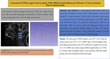
Assessment of pharyngeal airway space with MRI In oral submucous fibrosis: A cross-sectional observational study
Introduction
Oral submucous fibrosis (OSMF) alters the pharynx, which may affect airway size. MRI will be useful for diagnosing pharyngeal abnormalities. MRI is used to evaluate pharyngeal airway and soft palate changes in OSMF patients.
Materials and methods
This study is a cross-sectional observational study that included a sample size of 42 patients. Group A consisted of 21 patients with OSMF, while Group B consisted of 21 volunteers without OSMF, who served as the control group. The patients with OSMF were classified into Stages I, II, and III according to the categorization established by Pindborg JJ in 1989, Stop-Bang questionnaire was employed to assess obstructive sleep apnoea. Magnetic Resonance Imaging (MRI) was utilized to acquire evaluations of the pharyngeal airway, encompassing measurements in the midsagittal, cross-sectional width, length planes, and cross-sectional area with volume, for all participants. The Shapiro-Wilk test determines distribution normality. We utilized one-way ANOVA to compare the means between groups.
Results
The average age of OSMF patients was 45.9 ± 8.16, while the control group was 39.19 ± 4.21. Stage I of OSMF had the highest mean Stop Bang questionnaire score (2.75), followed by stage III (2.22), and stage II (1.75). Statistically significant differences (p < 0.001) were seen in volume, linear midsagittal planes, cross-sectional width and length planes, cross-sectional area, and soft palate breadth and length between OSMF and control groups.
Conclusion
MRI can effectively examine early changes in the pharyngeal airway of patients with OSMF thereby serving as a constructive diagnostic and motivational tool.
期刊介绍:
Journal of Oral Biology and Craniofacial Research (JOBCR)is the official journal of the Craniofacial Research Foundation (CRF). The journal aims to provide a common platform for both clinical and translational research and to promote interdisciplinary sciences in craniofacial region. JOBCR publishes content that includes diseases, injuries and defects in the head, neck, face, jaws and the hard and soft tissues of the mouth and jaws and face region; diagnosis and medical management of diseases specific to the orofacial tissues and of oral manifestations of systemic diseases; studies on identifying populations at risk of oral disease or in need of specific care, and comparing regional, environmental, social, and access similarities and differences in dental care between populations; diseases of the mouth and related structures like salivary glands, temporomandibular joints, facial muscles and perioral skin; biomedical engineering, tissue engineering and stem cells. The journal publishes reviews, commentaries, peer-reviewed original research articles, short communication, and case reports.

 求助内容:
求助内容: 应助结果提醒方式:


