Ala Al-Dubai, Mohammed Alhamed, Nurizzati Mohd Daud, Hak Yong Kim, Muhammad Mahadi Abdul Jamil, Syafiqah Saidin
{"title":"生物活性羟基磷灰石在聚多巴胺涂层上的成核 三维打印聚(乳酸)大孔支架用于骨移植应用","authors":"Ala Al-Dubai, Mohammed Alhamed, Nurizzati Mohd Daud, Hak Yong Kim, Muhammad Mahadi Abdul Jamil, Syafiqah Saidin","doi":"10.1007/s13369-024-09509-1","DOIUrl":null,"url":null,"abstract":"<div><p>The integration of poly (lactic acid) (PLA) in 3D printing has revolutionized the biomaterial engineering. This synergy between PLA and 3D printing holds immense potential for advancing tissue engineering and human health. The purpose of this study is to evaluate nucleated hydroxyapatite (HA) on polydopamine (PDA)-coated 3D printed PLA scaffolds, focusing on chemical composition, morphology, crystallinity, wettability, porosity, and biocompatibility via MTT assay. 3D printed PLA scaffolds were designed using computer-aided software (CAD). These scaffolds were immersed in a dopamine salt solution for 24 h to create a thin PDA layer and then placed in Simulated Body Fluid (SBF) for 5 days to stimulate apatite layer formation, observed through FE-SEM. PLA scaffolds had smooth surfaces, while PLA–PDA surfaces were different, and PLA–PDA–HA scaffolds revealed more homogeneous distribution of HA. PLA scaffolds had higher porosity (88%) and hydrophobic, whereas PLA–PDA scaffolds became hydrophilic due to the introduced amine and hydroxyl groups from the PDA coating. PLA–PDA scaffolds were amorphous, indicating reduced crystallinity, while PLA–PDA–HA displayed crystalline structure. The PLA–PDA–HA scaffolds declined most in weight loss (0.7%) due to the hydrolytic degradation of HA. In contrast the PLA–PDA scaffolds were the least degraded, with steady degradation trend lasting up to the 21st day of immersion. The PLA–PDA–HA scaffolds, with HA nucleation, exhibited the highest cell viability (145%) on day 7, emphasizing the crucial role of robust cell viability in tissue engineering. In conclusion, the PLA–PDA–HA scaffold was the most robust option due to its enhanced adhesion, biocompatibility, and potential protection against degradation.</p></div>","PeriodicalId":54354,"journal":{"name":"Arabian Journal for Science and Engineering","volume":"50 9","pages":"6523 - 6535"},"PeriodicalIF":2.6000,"publicationDate":"2024-08-28","publicationTypes":"Journal Article","fieldsOfStudy":null,"isOpenAccess":false,"openAccessPdf":"","citationCount":"0","resultStr":"{\"title\":\"Nucleation of Bioactive Hydroxyapatite on Polydopamine Coating Three-Dimensional Printed Poly (Lactic Acid) Macro-Porous Scaffold for Bone Grafting Application\",\"authors\":\"Ala Al-Dubai, Mohammed Alhamed, Nurizzati Mohd Daud, Hak Yong Kim, Muhammad Mahadi Abdul Jamil, Syafiqah Saidin\",\"doi\":\"10.1007/s13369-024-09509-1\",\"DOIUrl\":null,\"url\":null,\"abstract\":\"<div><p>The integration of poly (lactic acid) (PLA) in 3D printing has revolutionized the biomaterial engineering. This synergy between PLA and 3D printing holds immense potential for advancing tissue engineering and human health. The purpose of this study is to evaluate nucleated hydroxyapatite (HA) on polydopamine (PDA)-coated 3D printed PLA scaffolds, focusing on chemical composition, morphology, crystallinity, wettability, porosity, and biocompatibility via MTT assay. 3D printed PLA scaffolds were designed using computer-aided software (CAD). These scaffolds were immersed in a dopamine salt solution for 24 h to create a thin PDA layer and then placed in Simulated Body Fluid (SBF) for 5 days to stimulate apatite layer formation, observed through FE-SEM. PLA scaffolds had smooth surfaces, while PLA–PDA surfaces were different, and PLA–PDA–HA scaffolds revealed more homogeneous distribution of HA. PLA scaffolds had higher porosity (88%) and hydrophobic, whereas PLA–PDA scaffolds became hydrophilic due to the introduced amine and hydroxyl groups from the PDA coating. PLA–PDA scaffolds were amorphous, indicating reduced crystallinity, while PLA–PDA–HA displayed crystalline structure. The PLA–PDA–HA scaffolds declined most in weight loss (0.7%) due to the hydrolytic degradation of HA. In contrast the PLA–PDA scaffolds were the least degraded, with steady degradation trend lasting up to the 21st day of immersion. The PLA–PDA–HA scaffolds, with HA nucleation, exhibited the highest cell viability (145%) on day 7, emphasizing the crucial role of robust cell viability in tissue engineering. In conclusion, the PLA–PDA–HA scaffold was the most robust option due to its enhanced adhesion, biocompatibility, and potential protection against degradation.</p></div>\",\"PeriodicalId\":54354,\"journal\":{\"name\":\"Arabian Journal for Science and Engineering\",\"volume\":\"50 9\",\"pages\":\"6523 - 6535\"},\"PeriodicalIF\":2.6000,\"publicationDate\":\"2024-08-28\",\"publicationTypes\":\"Journal Article\",\"fieldsOfStudy\":null,\"isOpenAccess\":false,\"openAccessPdf\":\"\",\"citationCount\":\"0\",\"resultStr\":null,\"platform\":\"Semanticscholar\",\"paperid\":null,\"PeriodicalName\":\"Arabian Journal for Science and Engineering\",\"FirstCategoryId\":\"103\",\"ListUrlMain\":\"https://link.springer.com/article/10.1007/s13369-024-09509-1\",\"RegionNum\":4,\"RegionCategory\":\"综合性期刊\",\"ArticlePicture\":[],\"TitleCN\":null,\"AbstractTextCN\":null,\"PMCID\":null,\"EPubDate\":\"\",\"PubModel\":\"\",\"JCR\":\"Q2\",\"JCRName\":\"MULTIDISCIPLINARY SCIENCES\",\"Score\":null,\"Total\":0}","platform":"Semanticscholar","paperid":null,"PeriodicalName":"Arabian Journal for Science and Engineering","FirstCategoryId":"103","ListUrlMain":"https://link.springer.com/article/10.1007/s13369-024-09509-1","RegionNum":4,"RegionCategory":"综合性期刊","ArticlePicture":[],"TitleCN":null,"AbstractTextCN":null,"PMCID":null,"EPubDate":"","PubModel":"","JCR":"Q2","JCRName":"MULTIDISCIPLINARY SCIENCES","Score":null,"Total":0}
Nucleation of Bioactive Hydroxyapatite on Polydopamine Coating Three-Dimensional Printed Poly (Lactic Acid) Macro-Porous Scaffold for Bone Grafting Application
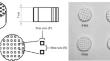
The integration of poly (lactic acid) (PLA) in 3D printing has revolutionized the biomaterial engineering. This synergy between PLA and 3D printing holds immense potential for advancing tissue engineering and human health. The purpose of this study is to evaluate nucleated hydroxyapatite (HA) on polydopamine (PDA)-coated 3D printed PLA scaffolds, focusing on chemical composition, morphology, crystallinity, wettability, porosity, and biocompatibility via MTT assay. 3D printed PLA scaffolds were designed using computer-aided software (CAD). These scaffolds were immersed in a dopamine salt solution for 24 h to create a thin PDA layer and then placed in Simulated Body Fluid (SBF) for 5 days to stimulate apatite layer formation, observed through FE-SEM. PLA scaffolds had smooth surfaces, while PLA–PDA surfaces were different, and PLA–PDA–HA scaffolds revealed more homogeneous distribution of HA. PLA scaffolds had higher porosity (88%) and hydrophobic, whereas PLA–PDA scaffolds became hydrophilic due to the introduced amine and hydroxyl groups from the PDA coating. PLA–PDA scaffolds were amorphous, indicating reduced crystallinity, while PLA–PDA–HA displayed crystalline structure. The PLA–PDA–HA scaffolds declined most in weight loss (0.7%) due to the hydrolytic degradation of HA. In contrast the PLA–PDA scaffolds were the least degraded, with steady degradation trend lasting up to the 21st day of immersion. The PLA–PDA–HA scaffolds, with HA nucleation, exhibited the highest cell viability (145%) on day 7, emphasizing the crucial role of robust cell viability in tissue engineering. In conclusion, the PLA–PDA–HA scaffold was the most robust option due to its enhanced adhesion, biocompatibility, and potential protection against degradation.
期刊介绍:
King Fahd University of Petroleum & Minerals (KFUPM) partnered with Springer to publish the Arabian Journal for Science and Engineering (AJSE).
AJSE, which has been published by KFUPM since 1975, is a recognized national, regional and international journal that provides a great opportunity for the dissemination of research advances from the Kingdom of Saudi Arabia, MENA and the world.
 求助内容:
求助内容: 应助结果提醒方式:


