Maura C. Eggink, Maarten J. F. de Wolf, Fenna A. Ebbens, Maartje M. L. de Win, Frederik G. Dikkers, Erik van Spronsen
{"title":"MRI-DWI 检测残余胆脂瘤:向最佳随访方案迈进","authors":"Maura C. Eggink, Maarten J. F. de Wolf, Fenna A. Ebbens, Maartje M. L. de Win, Frederik G. Dikkers, Erik van Spronsen","doi":"10.1007/s00405-024-08939-9","DOIUrl":null,"url":null,"abstract":"<h3 data-test=\"abstract-sub-heading\">Purpose</h3><p>To analyse diagnostic accuracy of MRI-DWI in detecting residual disease after cholesteatoma surgery and propose an optimum follow-up (FU) scheme.</p><h3 data-test=\"abstract-sub-heading\">Method</h3><p>A retrospective chart review of patients who had cholesteatoma surgery in a tertiary referral centre. 3.0 T non-echo planar diffusion weighted imaging was performed as part of routine FU or indicated on the basis of clinical suspicion of disease. Imaging outcome was verified per-operatively during a second-look procedure or ossicular chain reconstruction. Diagnostic parameters were calculated and stratified by FU length.</p><h3 data-test=\"abstract-sub-heading\">Results</h3><p>For the FU of 664 cholesteatoma surgeries, 1208 MRI-DWI were obtained and 235 second-look procedures were performed. Most MRI-DWI were obtained within 1.5 yrs of surgery. In this period, significantly less true positive MRI-DWI and significantly more false negative MRI-DWI for residual disease were found compared to other FU periods. Scanning after approximately 3 yrs yielded a significantly higher rate of true positive MRI-DWI, while sensitivity surpassed 80%. Younger patients had a higher risk of developing residual disease. Patients undergoing canal wall up surgery, as well as patients < 12 yrs, were at risk for false negative MRI-DWI. Obliteration reduces the risk of residual disease, while leading to less false negative MRI-DWI.</p><h3 data-test=\"abstract-sub-heading\">Conclusion</h3><p>A novel radiologic FU scheme for detecting residual disease is suggested for stable ears after cholesteatoma surgery: standard MRI-DWI approximately 3 and 5 yrs after primary surgery, as well as MRI-DWI after approximately 9 yrs for patients with specific risk factors (i.e., patients < 12 yrs or patients undergoing canal wall up surgery without obliteration).</p>","PeriodicalId":11952,"journal":{"name":"European Archives of Oto-Rhino-Laryngology","volume":"15 1","pages":""},"PeriodicalIF":1.9000,"publicationDate":"2024-09-13","publicationTypes":"Journal Article","fieldsOfStudy":null,"isOpenAccess":false,"openAccessPdf":"","citationCount":"0","resultStr":"{\"title\":\"MRI-DWI detection of residual cholesteatoma: moving toward an optimum follow-up scheme\",\"authors\":\"Maura C. Eggink, Maarten J. F. de Wolf, Fenna A. Ebbens, Maartje M. L. de Win, Frederik G. Dikkers, Erik van Spronsen\",\"doi\":\"10.1007/s00405-024-08939-9\",\"DOIUrl\":null,\"url\":null,\"abstract\":\"<h3 data-test=\\\"abstract-sub-heading\\\">Purpose</h3><p>To analyse diagnostic accuracy of MRI-DWI in detecting residual disease after cholesteatoma surgery and propose an optimum follow-up (FU) scheme.</p><h3 data-test=\\\"abstract-sub-heading\\\">Method</h3><p>A retrospective chart review of patients who had cholesteatoma surgery in a tertiary referral centre. 3.0 T non-echo planar diffusion weighted imaging was performed as part of routine FU or indicated on the basis of clinical suspicion of disease. Imaging outcome was verified per-operatively during a second-look procedure or ossicular chain reconstruction. Diagnostic parameters were calculated and stratified by FU length.</p><h3 data-test=\\\"abstract-sub-heading\\\">Results</h3><p>For the FU of 664 cholesteatoma surgeries, 1208 MRI-DWI were obtained and 235 second-look procedures were performed. Most MRI-DWI were obtained within 1.5 yrs of surgery. In this period, significantly less true positive MRI-DWI and significantly more false negative MRI-DWI for residual disease were found compared to other FU periods. Scanning after approximately 3 yrs yielded a significantly higher rate of true positive MRI-DWI, while sensitivity surpassed 80%. Younger patients had a higher risk of developing residual disease. Patients undergoing canal wall up surgery, as well as patients < 12 yrs, were at risk for false negative MRI-DWI. Obliteration reduces the risk of residual disease, while leading to less false negative MRI-DWI.</p><h3 data-test=\\\"abstract-sub-heading\\\">Conclusion</h3><p>A novel radiologic FU scheme for detecting residual disease is suggested for stable ears after cholesteatoma surgery: standard MRI-DWI approximately 3 and 5 yrs after primary surgery, as well as MRI-DWI after approximately 9 yrs for patients with specific risk factors (i.e., patients < 12 yrs or patients undergoing canal wall up surgery without obliteration).</p>\",\"PeriodicalId\":11952,\"journal\":{\"name\":\"European Archives of Oto-Rhino-Laryngology\",\"volume\":\"15 1\",\"pages\":\"\"},\"PeriodicalIF\":1.9000,\"publicationDate\":\"2024-09-13\",\"publicationTypes\":\"Journal Article\",\"fieldsOfStudy\":null,\"isOpenAccess\":false,\"openAccessPdf\":\"\",\"citationCount\":\"0\",\"resultStr\":null,\"platform\":\"Semanticscholar\",\"paperid\":null,\"PeriodicalName\":\"European Archives of Oto-Rhino-Laryngology\",\"FirstCategoryId\":\"3\",\"ListUrlMain\":\"https://doi.org/10.1007/s00405-024-08939-9\",\"RegionNum\":3,\"RegionCategory\":\"医学\",\"ArticlePicture\":[],\"TitleCN\":null,\"AbstractTextCN\":null,\"PMCID\":null,\"EPubDate\":\"\",\"PubModel\":\"\",\"JCR\":\"Q2\",\"JCRName\":\"OTORHINOLARYNGOLOGY\",\"Score\":null,\"Total\":0}","platform":"Semanticscholar","paperid":null,"PeriodicalName":"European Archives of Oto-Rhino-Laryngology","FirstCategoryId":"3","ListUrlMain":"https://doi.org/10.1007/s00405-024-08939-9","RegionNum":3,"RegionCategory":"医学","ArticlePicture":[],"TitleCN":null,"AbstractTextCN":null,"PMCID":null,"EPubDate":"","PubModel":"","JCR":"Q2","JCRName":"OTORHINOLARYNGOLOGY","Score":null,"Total":0}
MRI-DWI detection of residual cholesteatoma: moving toward an optimum follow-up scheme
Purpose
To analyse diagnostic accuracy of MRI-DWI in detecting residual disease after cholesteatoma surgery and propose an optimum follow-up (FU) scheme.
Method
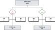
A retrospective chart review of patients who had cholesteatoma surgery in a tertiary referral centre. 3.0 T non-echo planar diffusion weighted imaging was performed as part of routine FU or indicated on the basis of clinical suspicion of disease. Imaging outcome was verified per-operatively during a second-look procedure or ossicular chain reconstruction. Diagnostic parameters were calculated and stratified by FU length.
Results
For the FU of 664 cholesteatoma surgeries, 1208 MRI-DWI were obtained and 235 second-look procedures were performed. Most MRI-DWI were obtained within 1.5 yrs of surgery. In this period, significantly less true positive MRI-DWI and significantly more false negative MRI-DWI for residual disease were found compared to other FU periods. Scanning after approximately 3 yrs yielded a significantly higher rate of true positive MRI-DWI, while sensitivity surpassed 80%. Younger patients had a higher risk of developing residual disease. Patients undergoing canal wall up surgery, as well as patients < 12 yrs, were at risk for false negative MRI-DWI. Obliteration reduces the risk of residual disease, while leading to less false negative MRI-DWI.
Conclusion
A novel radiologic FU scheme for detecting residual disease is suggested for stable ears after cholesteatoma surgery: standard MRI-DWI approximately 3 and 5 yrs after primary surgery, as well as MRI-DWI after approximately 9 yrs for patients with specific risk factors (i.e., patients < 12 yrs or patients undergoing canal wall up surgery without obliteration).
期刊介绍:
Official Journal of
European Union of Medical Specialists – ORL Section and Board
Official Journal of Confederation of European Oto-Rhino-Laryngology Head and Neck Surgery
"European Archives of Oto-Rhino-Laryngology" publishes original clinical reports and clinically relevant experimental studies, as well as short communications presenting new results of special interest. With peer review by a respected international editorial board and prompt English-language publication, the journal provides rapid dissemination of information by authors from around the world. This particular feature makes it the journal of choice for readers who want to be informed about the continuing state of the art concerning basic sciences and the diagnosis and management of diseases of the head and neck on an international level.
European Archives of Oto-Rhino-Laryngology was founded in 1864 as "Archiv für Ohrenheilkunde" by A. von Tröltsch, A. Politzer and H. Schwartze.
 求助内容:
求助内容: 应助结果提醒方式:


