Ana Mora-Boza, Adriana Mulero-Russe, Nikolas Di Caprio, Jason A. Burdick, Eric O’Neill, Ankur Singh, Andrés J. García
{"title":"水凝胶中可灌注微通道的简易光图案化,用于微生理系统","authors":"Ana Mora-Boza, Adriana Mulero-Russe, Nikolas Di Caprio, Jason A. Burdick, Eric O’Neill, Ankur Singh, Andrés J. García","doi":"10.1038/s41596-024-01041-8","DOIUrl":null,"url":null,"abstract":"<p>Perfusable hydrogels have garnered substantial attention in recent years for the fabrication of microphysiological systems. However, current methodologies to fabricate microchannels in hydrogel platforms involve sophisticated equipment and techniques, which hinder progress of the field. In this protocol, we present a cost-effective, simple, versatile and ultrafast method to create perfusable microchannels of complex shapes in photopolymerizable hydrogels. Our method uses one-step UV photocross-linking and a photomask printed on inexpensive transparent films, to photopattern both synthetic (PEG-norbornene) and natural (hyaluronic acid-norbornene) hydrogels in just 0.8 s. Moreover, these perfusable hydrogels are fully integrated into a custom-made microfluidic device that allows continuous fluid perfusion when connected to an external pump system. This methodology can be easily reproduced by professionals with basic laboratory skills and a fundamental knowledge of polymers and materials science. In this protocol, we demonstrate the functionality of our photopatterned hydrogels by seeding human endothelial cells into the microchannels, culturing them under dynamic conditions for 7 d, and exposing them to inflammatory stimuli to elicit cellular responses. This highlights the versatility of our platform in fabricating microphysiological systems and different microenvironments. The fabrication of perfusable channels within the hydrogels, including the fabrication of the microfluidic devices, requires ~3 d. The development of the cell-seeded microphysiological system, including the stimulation of cells, takes ~7 d. In conclusion, our approach provides a straightforward and widely applicable solution to simplify and reduce the cost of biofabrication techniques for developing functional in vitro models using perfusable three-dimensional hydrogels.</p>","PeriodicalId":18901,"journal":{"name":"Nature Protocols","volume":"19 1","pages":""},"PeriodicalIF":13.1000,"publicationDate":"2024-09-12","publicationTypes":"Journal Article","fieldsOfStudy":null,"isOpenAccess":false,"openAccessPdf":"","citationCount":"0","resultStr":"{\"title\":\"Facile photopatterning of perfusable microchannels in hydrogels for microphysiological systems\",\"authors\":\"Ana Mora-Boza, Adriana Mulero-Russe, Nikolas Di Caprio, Jason A. Burdick, Eric O’Neill, Ankur Singh, Andrés J. García\",\"doi\":\"10.1038/s41596-024-01041-8\",\"DOIUrl\":null,\"url\":null,\"abstract\":\"<p>Perfusable hydrogels have garnered substantial attention in recent years for the fabrication of microphysiological systems. However, current methodologies to fabricate microchannels in hydrogel platforms involve sophisticated equipment and techniques, which hinder progress of the field. In this protocol, we present a cost-effective, simple, versatile and ultrafast method to create perfusable microchannels of complex shapes in photopolymerizable hydrogels. Our method uses one-step UV photocross-linking and a photomask printed on inexpensive transparent films, to photopattern both synthetic (PEG-norbornene) and natural (hyaluronic acid-norbornene) hydrogels in just 0.8 s. Moreover, these perfusable hydrogels are fully integrated into a custom-made microfluidic device that allows continuous fluid perfusion when connected to an external pump system. This methodology can be easily reproduced by professionals with basic laboratory skills and a fundamental knowledge of polymers and materials science. In this protocol, we demonstrate the functionality of our photopatterned hydrogels by seeding human endothelial cells into the microchannels, culturing them under dynamic conditions for 7 d, and exposing them to inflammatory stimuli to elicit cellular responses. This highlights the versatility of our platform in fabricating microphysiological systems and different microenvironments. The fabrication of perfusable channels within the hydrogels, including the fabrication of the microfluidic devices, requires ~3 d. The development of the cell-seeded microphysiological system, including the stimulation of cells, takes ~7 d. In conclusion, our approach provides a straightforward and widely applicable solution to simplify and reduce the cost of biofabrication techniques for developing functional in vitro models using perfusable three-dimensional hydrogels.</p>\",\"PeriodicalId\":18901,\"journal\":{\"name\":\"Nature Protocols\",\"volume\":\"19 1\",\"pages\":\"\"},\"PeriodicalIF\":13.1000,\"publicationDate\":\"2024-09-12\",\"publicationTypes\":\"Journal Article\",\"fieldsOfStudy\":null,\"isOpenAccess\":false,\"openAccessPdf\":\"\",\"citationCount\":\"0\",\"resultStr\":null,\"platform\":\"Semanticscholar\",\"paperid\":null,\"PeriodicalName\":\"Nature Protocols\",\"FirstCategoryId\":\"99\",\"ListUrlMain\":\"https://doi.org/10.1038/s41596-024-01041-8\",\"RegionNum\":1,\"RegionCategory\":\"生物学\",\"ArticlePicture\":[],\"TitleCN\":null,\"AbstractTextCN\":null,\"PMCID\":null,\"EPubDate\":\"\",\"PubModel\":\"\",\"JCR\":\"Q1\",\"JCRName\":\"BIOCHEMICAL RESEARCH METHODS\",\"Score\":null,\"Total\":0}","platform":"Semanticscholar","paperid":null,"PeriodicalName":"Nature Protocols","FirstCategoryId":"99","ListUrlMain":"https://doi.org/10.1038/s41596-024-01041-8","RegionNum":1,"RegionCategory":"生物学","ArticlePicture":[],"TitleCN":null,"AbstractTextCN":null,"PMCID":null,"EPubDate":"","PubModel":"","JCR":"Q1","JCRName":"BIOCHEMICAL RESEARCH METHODS","Score":null,"Total":0}
Facile photopatterning of perfusable microchannels in hydrogels for microphysiological systems
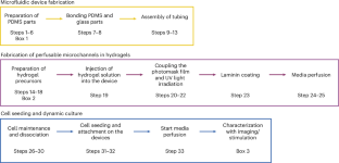
Perfusable hydrogels have garnered substantial attention in recent years for the fabrication of microphysiological systems. However, current methodologies to fabricate microchannels in hydrogel platforms involve sophisticated equipment and techniques, which hinder progress of the field. In this protocol, we present a cost-effective, simple, versatile and ultrafast method to create perfusable microchannels of complex shapes in photopolymerizable hydrogels. Our method uses one-step UV photocross-linking and a photomask printed on inexpensive transparent films, to photopattern both synthetic (PEG-norbornene) and natural (hyaluronic acid-norbornene) hydrogels in just 0.8 s. Moreover, these perfusable hydrogels are fully integrated into a custom-made microfluidic device that allows continuous fluid perfusion when connected to an external pump system. This methodology can be easily reproduced by professionals with basic laboratory skills and a fundamental knowledge of polymers and materials science. In this protocol, we demonstrate the functionality of our photopatterned hydrogels by seeding human endothelial cells into the microchannels, culturing them under dynamic conditions for 7 d, and exposing them to inflammatory stimuli to elicit cellular responses. This highlights the versatility of our platform in fabricating microphysiological systems and different microenvironments. The fabrication of perfusable channels within the hydrogels, including the fabrication of the microfluidic devices, requires ~3 d. The development of the cell-seeded microphysiological system, including the stimulation of cells, takes ~7 d. In conclusion, our approach provides a straightforward and widely applicable solution to simplify and reduce the cost of biofabrication techniques for developing functional in vitro models using perfusable three-dimensional hydrogels.
期刊介绍:
Nature Protocols focuses on publishing protocols used to address significant biological and biomedical science research questions, including methods grounded in physics and chemistry with practical applications to biological problems. The journal caters to a primary audience of research scientists and, as such, exclusively publishes protocols with research applications. Protocols primarily aimed at influencing patient management and treatment decisions are not featured.
The specific techniques covered encompass a wide range, including but not limited to: Biochemistry, Cell biology, Cell culture, Chemical modification, Computational biology, Developmental biology, Epigenomics, Genetic analysis, Genetic modification, Genomics, Imaging, Immunology, Isolation, purification, and separation, Lipidomics, Metabolomics, Microbiology, Model organisms, Nanotechnology, Neuroscience, Nucleic-acid-based molecular biology, Pharmacology, Plant biology, Protein analysis, Proteomics, Spectroscopy, Structural biology, Synthetic chemistry, Tissue culture, Toxicology, and Virology.