Jae Young Joo, Dallah Yoo, Jae-Myoung Kim, Chaewon Shin, Tae-Beom Ahn
{"title":"体位改变对帕金森病伴正位性低血压患者脑灌注的影响","authors":"Jae Young Joo, Dallah Yoo, Jae-Myoung Kim, Chaewon Shin, Tae-Beom Ahn","doi":"10.14802/jmd.24104","DOIUrl":null,"url":null,"abstract":"<p><strong>Objective: </strong>Orthostatic hypotension (OH) is one of the most common autonomic dysfunctions in Parkinson's disease (PD) patients. However, many patients with OH are asymptomatic. Conversely, orthostatic dizziness (OD) is not always associated with OH. We investigated the effects of positional changes on cerebral perfusion in patients with PD and OH.</p><p><strong>Methods: </strong>We enrolled 42 patients, comprising 31 PD patients and 11 healthy controls. All the subjects underwent the following clinical assessments: the OH questionnaire, head-up tilt test (HUTT) with transcranial Doppler (TCD), near-infrared spectroscopy, measurement of the change in oxygenated hemoglobin (ΔHboxy) during the squat-to-stand test (SST), measurement of the time derivative of total hemoglobin (DHbtot), and time taken to reach the peak (peak time [PT]) of DHbtot after restanding.</p><p><strong>Results: </strong>The mean flow velocity change (ΔMFV) in the TCD during the HUTT failed to differentiate between the PD-OH(+) and PD-OH(-) groups. The change in oxygenated hemoglobin ΔHboxy was greater in the PD-OH(+) group, which persisted for 9 min until the end of the HUTT only in the left hemisphere. During SST, PT was significantly delayed in the left hemisphere in PD-OH(+) patients.</p><p><strong>Conclusion: </strong>Although TCD demonstrated no significant difference in ΔMFV, the parameters measured by near-infrared spectroscopy, such as ΔHboxy during HUTT and PT during the SST, significantly increased ΔHboxy or delayed PT in the left hemisphere of PD-OH(+). Positional changes have a detrimental effect on cerebral hemodynamics in patients with PD and OH, especially in the left hemisphere.</p>","PeriodicalId":16372,"journal":{"name":"Journal of Movement Disorders","volume":" ","pages":"408-415"},"PeriodicalIF":2.8000,"publicationDate":"2024-10-01","publicationTypes":"Journal Article","fieldsOfStudy":null,"isOpenAccess":false,"openAccessPdf":"https://www.ncbi.nlm.nih.gov/pmc/articles/PMC11540534/pdf/","citationCount":"0","resultStr":"{\"title\":\"Effect of Positional Changes on Cerebral Perfusion in Parkinson's Disease Patients With Orthostatic Hypotension.\",\"authors\":\"Jae Young Joo, Dallah Yoo, Jae-Myoung Kim, Chaewon Shin, Tae-Beom Ahn\",\"doi\":\"10.14802/jmd.24104\",\"DOIUrl\":null,\"url\":null,\"abstract\":\"<p><strong>Objective: </strong>Orthostatic hypotension (OH) is one of the most common autonomic dysfunctions in Parkinson's disease (PD) patients. However, many patients with OH are asymptomatic. Conversely, orthostatic dizziness (OD) is not always associated with OH. We investigated the effects of positional changes on cerebral perfusion in patients with PD and OH.</p><p><strong>Methods: </strong>We enrolled 42 patients, comprising 31 PD patients and 11 healthy controls. All the subjects underwent the following clinical assessments: the OH questionnaire, head-up tilt test (HUTT) with transcranial Doppler (TCD), near-infrared spectroscopy, measurement of the change in oxygenated hemoglobin (ΔHboxy) during the squat-to-stand test (SST), measurement of the time derivative of total hemoglobin (DHbtot), and time taken to reach the peak (peak time [PT]) of DHbtot after restanding.</p><p><strong>Results: </strong>The mean flow velocity change (ΔMFV) in the TCD during the HUTT failed to differentiate between the PD-OH(+) and PD-OH(-) groups. The change in oxygenated hemoglobin ΔHboxy was greater in the PD-OH(+) group, which persisted for 9 min until the end of the HUTT only in the left hemisphere. During SST, PT was significantly delayed in the left hemisphere in PD-OH(+) patients.</p><p><strong>Conclusion: </strong>Although TCD demonstrated no significant difference in ΔMFV, the parameters measured by near-infrared spectroscopy, such as ΔHboxy during HUTT and PT during the SST, significantly increased ΔHboxy or delayed PT in the left hemisphere of PD-OH(+). Positional changes have a detrimental effect on cerebral hemodynamics in patients with PD and OH, especially in the left hemisphere.</p>\",\"PeriodicalId\":16372,\"journal\":{\"name\":\"Journal of Movement Disorders\",\"volume\":\" \",\"pages\":\"408-415\"},\"PeriodicalIF\":2.8000,\"publicationDate\":\"2024-10-01\",\"publicationTypes\":\"Journal Article\",\"fieldsOfStudy\":null,\"isOpenAccess\":false,\"openAccessPdf\":\"https://www.ncbi.nlm.nih.gov/pmc/articles/PMC11540534/pdf/\",\"citationCount\":\"0\",\"resultStr\":null,\"platform\":\"Semanticscholar\",\"paperid\":null,\"PeriodicalName\":\"Journal of Movement Disorders\",\"FirstCategoryId\":\"3\",\"ListUrlMain\":\"https://doi.org/10.14802/jmd.24104\",\"RegionNum\":4,\"RegionCategory\":\"医学\",\"ArticlePicture\":[],\"TitleCN\":null,\"AbstractTextCN\":null,\"PMCID\":null,\"EPubDate\":\"2024/9/9 0:00:00\",\"PubModel\":\"Epub\",\"JCR\":\"Q2\",\"JCRName\":\"CLINICAL NEUROLOGY\",\"Score\":null,\"Total\":0}","platform":"Semanticscholar","paperid":null,"PeriodicalName":"Journal of Movement Disorders","FirstCategoryId":"3","ListUrlMain":"https://doi.org/10.14802/jmd.24104","RegionNum":4,"RegionCategory":"医学","ArticlePicture":[],"TitleCN":null,"AbstractTextCN":null,"PMCID":null,"EPubDate":"2024/9/9 0:00:00","PubModel":"Epub","JCR":"Q2","JCRName":"CLINICAL NEUROLOGY","Score":null,"Total":0}
Effect of Positional Changes on Cerebral Perfusion in Parkinson's Disease Patients With Orthostatic Hypotension.
Objective: Orthostatic hypotension (OH) is one of the most common autonomic dysfunctions in Parkinson's disease (PD) patients. However, many patients with OH are asymptomatic. Conversely, orthostatic dizziness (OD) is not always associated with OH. We investigated the effects of positional changes on cerebral perfusion in patients with PD and OH.
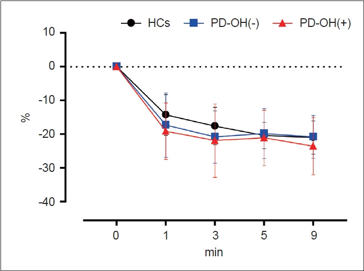
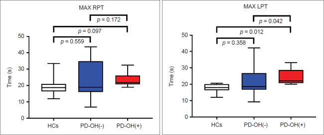
Methods: We enrolled 42 patients, comprising 31 PD patients and 11 healthy controls. All the subjects underwent the following clinical assessments: the OH questionnaire, head-up tilt test (HUTT) with transcranial Doppler (TCD), near-infrared spectroscopy, measurement of the change in oxygenated hemoglobin (ΔHboxy) during the squat-to-stand test (SST), measurement of the time derivative of total hemoglobin (DHbtot), and time taken to reach the peak (peak time [PT]) of DHbtot after restanding.
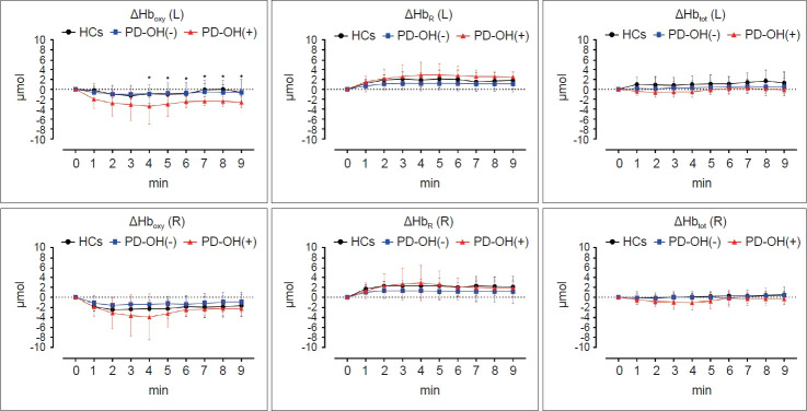
Results: The mean flow velocity change (ΔMFV) in the TCD during the HUTT failed to differentiate between the PD-OH(+) and PD-OH(-) groups. The change in oxygenated hemoglobin ΔHboxy was greater in the PD-OH(+) group, which persisted for 9 min until the end of the HUTT only in the left hemisphere. During SST, PT was significantly delayed in the left hemisphere in PD-OH(+) patients.
Conclusion: Although TCD demonstrated no significant difference in ΔMFV, the parameters measured by near-infrared spectroscopy, such as ΔHboxy during HUTT and PT during the SST, significantly increased ΔHboxy or delayed PT in the left hemisphere of PD-OH(+). Positional changes have a detrimental effect on cerebral hemodynamics in patients with PD and OH, especially in the left hemisphere.
 求助内容:
求助内容: 应助结果提醒方式:


