J. Soukup , M. Zelená , F. Weisz , M. Kostelanská , E. Nohýnková , P. Tůmová
{"title":"利用膨胀显微镜对贾第虫肠道细胞组织进行成像,发现非典型中心蛋白定位。","authors":"J. Soukup , M. Zelená , F. Weisz , M. Kostelanská , E. Nohýnková , P. Tůmová","doi":"10.1016/j.exppara.2024.108831","DOIUrl":null,"url":null,"abstract":"<div><p>Advanced imaging of microorganisms, including protists, is challenging due to their small size. Specimen expansion prior to imaging is thus beneficial to increase resolution and cellular details. Here, we present a sample preparation workflow for improved observations of the single-celled eukaryotic pathogen <em>Giardia intestinalis</em> (Excavata, Metamonada). The binucleated trophozoites colonize the small intestine of humans and animals and cause a diarrhoeal disease. Their remarkable morphology includes two nuclei and a pronounced microtubular cytoskeleton enabling cell motility, attachment and proliferation. By use of expansion and confocal microscopy, we resolved in a great detail subcellular structures and organelles of the parasite cell. The acquired spatial resolution enabled novel observations of centrin localization at <em>Giardia</em> basal bodies. Interestingly, non-luminal centrin localization between the <em>Giardia</em> basal bodies was observed, which is an atypical eukaryotic arrangement. Our protocol includes antibody staining and can be used for the localization of epitope-tagged proteins, as well as for differential organelle labelling by amino reactive esters. This fast and simple technique is suitable for routine use without a superresolution microscopy equipment.</p></div>","PeriodicalId":12117,"journal":{"name":"Experimental parasitology","volume":"266 ","pages":"Article 108831"},"PeriodicalIF":1.6000,"publicationDate":"2024-09-06","publicationTypes":"Journal Article","fieldsOfStudy":null,"isOpenAccess":false,"openAccessPdf":"https://www.sciencedirect.com/science/article/pii/S0014489424001346/pdfft?md5=707dd25dc64bd200a45705c2b5c417d8&pid=1-s2.0-S0014489424001346-main.pdf","citationCount":"0","resultStr":"{\"title\":\"Imaging Giardia intestinalis cellular organisation using expansion microscopy reveals atypical centrin localisation\",\"authors\":\"J. Soukup , M. Zelená , F. Weisz , M. Kostelanská , E. Nohýnková , P. Tůmová\",\"doi\":\"10.1016/j.exppara.2024.108831\",\"DOIUrl\":null,\"url\":null,\"abstract\":\"<div><p>Advanced imaging of microorganisms, including protists, is challenging due to their small size. Specimen expansion prior to imaging is thus beneficial to increase resolution and cellular details. Here, we present a sample preparation workflow for improved observations of the single-celled eukaryotic pathogen <em>Giardia intestinalis</em> (Excavata, Metamonada). The binucleated trophozoites colonize the small intestine of humans and animals and cause a diarrhoeal disease. Their remarkable morphology includes two nuclei and a pronounced microtubular cytoskeleton enabling cell motility, attachment and proliferation. By use of expansion and confocal microscopy, we resolved in a great detail subcellular structures and organelles of the parasite cell. The acquired spatial resolution enabled novel observations of centrin localization at <em>Giardia</em> basal bodies. Interestingly, non-luminal centrin localization between the <em>Giardia</em> basal bodies was observed, which is an atypical eukaryotic arrangement. Our protocol includes antibody staining and can be used for the localization of epitope-tagged proteins, as well as for differential organelle labelling by amino reactive esters. This fast and simple technique is suitable for routine use without a superresolution microscopy equipment.</p></div>\",\"PeriodicalId\":12117,\"journal\":{\"name\":\"Experimental parasitology\",\"volume\":\"266 \",\"pages\":\"Article 108831\"},\"PeriodicalIF\":1.6000,\"publicationDate\":\"2024-09-06\",\"publicationTypes\":\"Journal Article\",\"fieldsOfStudy\":null,\"isOpenAccess\":false,\"openAccessPdf\":\"https://www.sciencedirect.com/science/article/pii/S0014489424001346/pdfft?md5=707dd25dc64bd200a45705c2b5c417d8&pid=1-s2.0-S0014489424001346-main.pdf\",\"citationCount\":\"0\",\"resultStr\":null,\"platform\":\"Semanticscholar\",\"paperid\":null,\"PeriodicalName\":\"Experimental parasitology\",\"FirstCategoryId\":\"3\",\"ListUrlMain\":\"https://www.sciencedirect.com/science/article/pii/S0014489424001346\",\"RegionNum\":4,\"RegionCategory\":\"医学\",\"ArticlePicture\":[],\"TitleCN\":null,\"AbstractTextCN\":null,\"PMCID\":null,\"EPubDate\":\"\",\"PubModel\":\"\",\"JCR\":\"Q3\",\"JCRName\":\"PARASITOLOGY\",\"Score\":null,\"Total\":0}","platform":"Semanticscholar","paperid":null,"PeriodicalName":"Experimental parasitology","FirstCategoryId":"3","ListUrlMain":"https://www.sciencedirect.com/science/article/pii/S0014489424001346","RegionNum":4,"RegionCategory":"医学","ArticlePicture":[],"TitleCN":null,"AbstractTextCN":null,"PMCID":null,"EPubDate":"","PubModel":"","JCR":"Q3","JCRName":"PARASITOLOGY","Score":null,"Total":0}
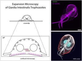
Advanced imaging of microorganisms, including protists, is challenging due to their small size. Specimen expansion prior to imaging is thus beneficial to increase resolution and cellular details. Here, we present a sample preparation workflow for improved observations of the single-celled eukaryotic pathogen Giardia intestinalis (Excavata, Metamonada). The binucleated trophozoites colonize the small intestine of humans and animals and cause a diarrhoeal disease. Their remarkable morphology includes two nuclei and a pronounced microtubular cytoskeleton enabling cell motility, attachment and proliferation. By use of expansion and confocal microscopy, we resolved in a great detail subcellular structures and organelles of the parasite cell. The acquired spatial resolution enabled novel observations of centrin localization at Giardia basal bodies. Interestingly, non-luminal centrin localization between the Giardia basal bodies was observed, which is an atypical eukaryotic arrangement. Our protocol includes antibody staining and can be used for the localization of epitope-tagged proteins, as well as for differential organelle labelling by amino reactive esters. This fast and simple technique is suitable for routine use without a superresolution microscopy equipment.
期刊介绍:
Experimental Parasitology emphasizes modern approaches to parasitology, including molecular biology and immunology. The journal features original research papers on the physiological, metabolic, immunologic, biochemical, nutritional, and chemotherapeutic aspects of parasites and host-parasite relationships.
 求助内容:
求助内容: 应助结果提醒方式:


