Robert Graf, Paul-Sören Platzek, Evamaria Olga Riedel, Su Hwan Kim, Nicolas Lenhart, Constanze Ramschütz, Karolin Johanna Paprottka, Olivia Ruriko Kertels, Hendrik Kristian Möller, Matan Atad, Robin Bülow, Nicole Werner, Henry Völzke, Carsten Oliver Schmidt, Benedikt Wiestler, Johannes C Paetzold, Daniel Rueckert, Jan Stefan Kirschke
{"title":"根据 T2w FSE 和低分辨率轴向 Dixon 合成高分辨率脊柱 STIR 和 T1w 图像。","authors":"Robert Graf, Paul-Sören Platzek, Evamaria Olga Riedel, Su Hwan Kim, Nicolas Lenhart, Constanze Ramschütz, Karolin Johanna Paprottka, Olivia Ruriko Kertels, Hendrik Kristian Möller, Matan Atad, Robin Bülow, Nicole Werner, Henry Völzke, Carsten Oliver Schmidt, Benedikt Wiestler, Johannes C Paetzold, Daniel Rueckert, Jan Stefan Kirschke","doi":"10.1007/s00330-024-11047-1","DOIUrl":null,"url":null,"abstract":"<p><strong>Objectives: </strong>To generate sagittal T1-weighted fast spin echo (T1w FSE) and short tau inversion recovery (STIR) images from sagittal T2-weighted (T2w) FSE and axial T1w gradient echo Dixon technique (T1w-Dixon) sequences.</p><p><strong>Materials and methods: </strong>This retrospective study used three existing datasets: \"Study of Health in Pomerania\" (SHIP, 3142 subjects, 1.5 Tesla), \"German National Cohort\" (NAKO, 2000 subjects, 3 Tesla), and an internal dataset (157 patients 1.5/3 Tesla). We generated synthetic sagittal T1w FSE and STIR images from sagittal T2w FSE and low-resolution axial T1w-Dixon sequences based on two successively applied 3D Pix2Pix deep learning models. \"Peak signal-to-noise ratio\" (PSNR) and \"structural similarity index metric\" (SSIM) were used to evaluate the generated image quality on an ablations test. A Turing test, where seven radiologists rated 240 images as either natively acquired or generated, was evaluated using misclassification rate and Fleiss kappa interrater agreement.</p><p><strong>Results: </strong>Including axial T1w-Dixon or T1w FSE images resulted in higher image quality in generated T1w FSE (PSNR = 26.942, SSIM = 0.965) and STIR (PSNR = 28.86, SSIM = 0.948) images compared to using only single T2w images as input (PSNR = 23.076/24.677 SSIM = 0.952/0.928). Radiologists had difficulty identifying generated images (misclassification rate: 0.39 ± 0.09 for T1w FSE, 0.42 ± 0.18 for STIR) and showed low interrater agreement on suspicious images (Fleiss kappa: 0.09 for T1w/STIR).</p><p><strong>Conclusions: </strong>Axial T1w-Dixon and sagittal T2w FSE images contain sufficient information to generate sagittal T1w FSE and STIR images.</p><p><strong>Clinical relevance statement: </strong>T1w fast spin echo and short tau inversion recovery can be retroactively added to existing datasets, saving MRI time and enabling retrospective analysis, such as evaluating bone marrow pathologies.</p><p><strong>Key points: </strong>Sagittal T2-weighted images alone were insufficient for differentiating fat and water and to generate T1-weighted images. Axial T1w Dixon technique, together with a T2-weighted sequence, produced realistic sagittal T1-weighted images. Our approach can be used to retrospectively generate STIR and T1-weighted fast spin echo sequences.</p>","PeriodicalId":12076,"journal":{"name":"European Radiology","volume":" ","pages":"1761-1771"},"PeriodicalIF":4.7000,"publicationDate":"2025-04-01","publicationTypes":"Journal Article","fieldsOfStudy":null,"isOpenAccess":false,"openAccessPdf":"https://www.ncbi.nlm.nih.gov/pmc/articles/PMC11913981/pdf/","citationCount":"0","resultStr":"{\"title\":\"Generating synthetic high-resolution spinal STIR and T1w images from T2w FSE and low-resolution axial Dixon.\",\"authors\":\"Robert Graf, Paul-Sören Platzek, Evamaria Olga Riedel, Su Hwan Kim, Nicolas Lenhart, Constanze Ramschütz, Karolin Johanna Paprottka, Olivia Ruriko Kertels, Hendrik Kristian Möller, Matan Atad, Robin Bülow, Nicole Werner, Henry Völzke, Carsten Oliver Schmidt, Benedikt Wiestler, Johannes C Paetzold, Daniel Rueckert, Jan Stefan Kirschke\",\"doi\":\"10.1007/s00330-024-11047-1\",\"DOIUrl\":null,\"url\":null,\"abstract\":\"<p><strong>Objectives: </strong>To generate sagittal T1-weighted fast spin echo (T1w FSE) and short tau inversion recovery (STIR) images from sagittal T2-weighted (T2w) FSE and axial T1w gradient echo Dixon technique (T1w-Dixon) sequences.</p><p><strong>Materials and methods: </strong>This retrospective study used three existing datasets: \\\"Study of Health in Pomerania\\\" (SHIP, 3142 subjects, 1.5 Tesla), \\\"German National Cohort\\\" (NAKO, 2000 subjects, 3 Tesla), and an internal dataset (157 patients 1.5/3 Tesla). We generated synthetic sagittal T1w FSE and STIR images from sagittal T2w FSE and low-resolution axial T1w-Dixon sequences based on two successively applied 3D Pix2Pix deep learning models. \\\"Peak signal-to-noise ratio\\\" (PSNR) and \\\"structural similarity index metric\\\" (SSIM) were used to evaluate the generated image quality on an ablations test. A Turing test, where seven radiologists rated 240 images as either natively acquired or generated, was evaluated using misclassification rate and Fleiss kappa interrater agreement.</p><p><strong>Results: </strong>Including axial T1w-Dixon or T1w FSE images resulted in higher image quality in generated T1w FSE (PSNR = 26.942, SSIM = 0.965) and STIR (PSNR = 28.86, SSIM = 0.948) images compared to using only single T2w images as input (PSNR = 23.076/24.677 SSIM = 0.952/0.928). Radiologists had difficulty identifying generated images (misclassification rate: 0.39 ± 0.09 for T1w FSE, 0.42 ± 0.18 for STIR) and showed low interrater agreement on suspicious images (Fleiss kappa: 0.09 for T1w/STIR).</p><p><strong>Conclusions: </strong>Axial T1w-Dixon and sagittal T2w FSE images contain sufficient information to generate sagittal T1w FSE and STIR images.</p><p><strong>Clinical relevance statement: </strong>T1w fast spin echo and short tau inversion recovery can be retroactively added to existing datasets, saving MRI time and enabling retrospective analysis, such as evaluating bone marrow pathologies.</p><p><strong>Key points: </strong>Sagittal T2-weighted images alone were insufficient for differentiating fat and water and to generate T1-weighted images. Axial T1w Dixon technique, together with a T2-weighted sequence, produced realistic sagittal T1-weighted images. Our approach can be used to retrospectively generate STIR and T1-weighted fast spin echo sequences.</p>\",\"PeriodicalId\":12076,\"journal\":{\"name\":\"European Radiology\",\"volume\":\" \",\"pages\":\"1761-1771\"},\"PeriodicalIF\":4.7000,\"publicationDate\":\"2025-04-01\",\"publicationTypes\":\"Journal Article\",\"fieldsOfStudy\":null,\"isOpenAccess\":false,\"openAccessPdf\":\"https://www.ncbi.nlm.nih.gov/pmc/articles/PMC11913981/pdf/\",\"citationCount\":\"0\",\"resultStr\":null,\"platform\":\"Semanticscholar\",\"paperid\":null,\"PeriodicalName\":\"European Radiology\",\"FirstCategoryId\":\"3\",\"ListUrlMain\":\"https://doi.org/10.1007/s00330-024-11047-1\",\"RegionNum\":2,\"RegionCategory\":\"医学\",\"ArticlePicture\":[],\"TitleCN\":null,\"AbstractTextCN\":null,\"PMCID\":null,\"EPubDate\":\"2024/9/4 0:00:00\",\"PubModel\":\"Epub\",\"JCR\":\"Q1\",\"JCRName\":\"RADIOLOGY, NUCLEAR MEDICINE & MEDICAL IMAGING\",\"Score\":null,\"Total\":0}","platform":"Semanticscholar","paperid":null,"PeriodicalName":"European Radiology","FirstCategoryId":"3","ListUrlMain":"https://doi.org/10.1007/s00330-024-11047-1","RegionNum":2,"RegionCategory":"医学","ArticlePicture":[],"TitleCN":null,"AbstractTextCN":null,"PMCID":null,"EPubDate":"2024/9/4 0:00:00","PubModel":"Epub","JCR":"Q1","JCRName":"RADIOLOGY, NUCLEAR MEDICINE & MEDICAL IMAGING","Score":null,"Total":0}
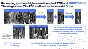
Generating synthetic high-resolution spinal STIR and T1w images from T2w FSE and low-resolution axial Dixon.
Objectives: To generate sagittal T1-weighted fast spin echo (T1w FSE) and short tau inversion recovery (STIR) images from sagittal T2-weighted (T2w) FSE and axial T1w gradient echo Dixon technique (T1w-Dixon) sequences.
Materials and methods: This retrospective study used three existing datasets: "Study of Health in Pomerania" (SHIP, 3142 subjects, 1.5 Tesla), "German National Cohort" (NAKO, 2000 subjects, 3 Tesla), and an internal dataset (157 patients 1.5/3 Tesla). We generated synthetic sagittal T1w FSE and STIR images from sagittal T2w FSE and low-resolution axial T1w-Dixon sequences based on two successively applied 3D Pix2Pix deep learning models. "Peak signal-to-noise ratio" (PSNR) and "structural similarity index metric" (SSIM) were used to evaluate the generated image quality on an ablations test. A Turing test, where seven radiologists rated 240 images as either natively acquired or generated, was evaluated using misclassification rate and Fleiss kappa interrater agreement.
Results: Including axial T1w-Dixon or T1w FSE images resulted in higher image quality in generated T1w FSE (PSNR = 26.942, SSIM = 0.965) and STIR (PSNR = 28.86, SSIM = 0.948) images compared to using only single T2w images as input (PSNR = 23.076/24.677 SSIM = 0.952/0.928). Radiologists had difficulty identifying generated images (misclassification rate: 0.39 ± 0.09 for T1w FSE, 0.42 ± 0.18 for STIR) and showed low interrater agreement on suspicious images (Fleiss kappa: 0.09 for T1w/STIR).
Conclusions: Axial T1w-Dixon and sagittal T2w FSE images contain sufficient information to generate sagittal T1w FSE and STIR images.
Clinical relevance statement: T1w fast spin echo and short tau inversion recovery can be retroactively added to existing datasets, saving MRI time and enabling retrospective analysis, such as evaluating bone marrow pathologies.
Key points: Sagittal T2-weighted images alone were insufficient for differentiating fat and water and to generate T1-weighted images. Axial T1w Dixon technique, together with a T2-weighted sequence, produced realistic sagittal T1-weighted images. Our approach can be used to retrospectively generate STIR and T1-weighted fast spin echo sequences.
期刊介绍:
European Radiology (ER) continuously updates scientific knowledge in radiology by publication of strong original articles and state-of-the-art reviews written by leading radiologists. A well balanced combination of review articles, original papers, short communications from European radiological congresses and information on society matters makes ER an indispensable source for current information in this field.
This is the Journal of the European Society of Radiology, and the official journal of a number of societies.
From 2004-2008 supplements to European Radiology were published under its companion, European Radiology Supplements, ISSN 1613-3749.

 求助内容:
求助内容: 应助结果提醒方式:


