{"title":"人工智能辅助自动筛查来自不同扫描仪的计算机断层扫描图像中的机会性骨质疏松症。","authors":"Yan Wu, Xiaopeng Yang, Mingyue Wang, Yanbang Lian, Ping Hou, Xiangfei Chai, Qiong Dai, Baoxin Qian, Yaojun Jiang, Jianbo Gao","doi":"10.1007/s00330-024-11046-2","DOIUrl":null,"url":null,"abstract":"<p><strong>Objectives: </strong>It is feasible to evaluate bone mineral density (BMD) and detect osteoporosis through an artificial intelligence (AI)-assisted system by using quantitative computed tomography (QCT) as a reference without additional radiation exposure or cost.</p><p><strong>Methods: </strong>A deep-learning model developed based on 3312 low-dose chest computed tomography (LDCT) scans (trained with 2337 and tested with 975) achieved a mean dice similarity coefficient of 95.8% for T1-T12, L1, and L2 vertebral body (VB) segmentation on test data. We performed a model evaluation based on 4401 LDCT scans (obtained from scanners of 3 different manufacturers as external validation data). The BMD values of all individuals were extracted from three consecutive VBs: T12 to L2. Line regression and Bland‒Altman analyses were used to evaluate the overall detection performance. Sensitivity and specificity were used to evaluate the diagnostic performance for normal, osteopenia, and osteoporosis patients.</p><p><strong>Results: </strong>Compared with the QCT results as the diagnostic standard, the BMD assessed had a mean error of (- 0.28, 2.37) mg/cm<sup>3</sup>. Overall, the sensitivity of a normal diagnosis was greater than that of a diagnosis of osteopenia or osteoporosis. For the diagnosis of osteoporosis, the model achieved a sensitivity > 86% and a specificity > 98%.</p><p><strong>Conclusion: </strong>The developed tool is clinically applicable and helpful for the positioning and analysis of VBs, the measurement of BMD, and the screening of osteopenia and osteoporosis.</p><p><strong>Clinical relevance statement: </strong>The developed system achieved high accuracy for automatic opportunistic osteoporosis screening using low-dose chest CT scans and performed well on CT images collected from different scanners.</p><p><strong>Key points: </strong>Osteoporosis is a prevalent but underdiagnosed condition that can increase the risk of fractures. This system could automatically and opportunistically screen for osteoporosis using low-dose chest CT scans obtained for lung cancer screening. The developed system performed well on CT images collected from different scanners and did not differ with patient age or sex.</p>","PeriodicalId":12076,"journal":{"name":"European Radiology","volume":" ","pages":"2287-2295"},"PeriodicalIF":4.7000,"publicationDate":"2025-04-01","publicationTypes":"Journal Article","fieldsOfStudy":null,"isOpenAccess":false,"openAccessPdf":"","citationCount":"0","resultStr":"{\"title\":\"Artificial intelligence assisted automatic screening of opportunistic osteoporosis in computed tomography images from different scanners.\",\"authors\":\"Yan Wu, Xiaopeng Yang, Mingyue Wang, Yanbang Lian, Ping Hou, Xiangfei Chai, Qiong Dai, Baoxin Qian, Yaojun Jiang, Jianbo Gao\",\"doi\":\"10.1007/s00330-024-11046-2\",\"DOIUrl\":null,\"url\":null,\"abstract\":\"<p><strong>Objectives: </strong>It is feasible to evaluate bone mineral density (BMD) and detect osteoporosis through an artificial intelligence (AI)-assisted system by using quantitative computed tomography (QCT) as a reference without additional radiation exposure or cost.</p><p><strong>Methods: </strong>A deep-learning model developed based on 3312 low-dose chest computed tomography (LDCT) scans (trained with 2337 and tested with 975) achieved a mean dice similarity coefficient of 95.8% for T1-T12, L1, and L2 vertebral body (VB) segmentation on test data. We performed a model evaluation based on 4401 LDCT scans (obtained from scanners of 3 different manufacturers as external validation data). The BMD values of all individuals were extracted from three consecutive VBs: T12 to L2. Line regression and Bland‒Altman analyses were used to evaluate the overall detection performance. Sensitivity and specificity were used to evaluate the diagnostic performance for normal, osteopenia, and osteoporosis patients.</p><p><strong>Results: </strong>Compared with the QCT results as the diagnostic standard, the BMD assessed had a mean error of (- 0.28, 2.37) mg/cm<sup>3</sup>. Overall, the sensitivity of a normal diagnosis was greater than that of a diagnosis of osteopenia or osteoporosis. For the diagnosis of osteoporosis, the model achieved a sensitivity > 86% and a specificity > 98%.</p><p><strong>Conclusion: </strong>The developed tool is clinically applicable and helpful for the positioning and analysis of VBs, the measurement of BMD, and the screening of osteopenia and osteoporosis.</p><p><strong>Clinical relevance statement: </strong>The developed system achieved high accuracy for automatic opportunistic osteoporosis screening using low-dose chest CT scans and performed well on CT images collected from different scanners.</p><p><strong>Key points: </strong>Osteoporosis is a prevalent but underdiagnosed condition that can increase the risk of fractures. This system could automatically and opportunistically screen for osteoporosis using low-dose chest CT scans obtained for lung cancer screening. The developed system performed well on CT images collected from different scanners and did not differ with patient age or sex.</p>\",\"PeriodicalId\":12076,\"journal\":{\"name\":\"European Radiology\",\"volume\":\" \",\"pages\":\"2287-2295\"},\"PeriodicalIF\":4.7000,\"publicationDate\":\"2025-04-01\",\"publicationTypes\":\"Journal Article\",\"fieldsOfStudy\":null,\"isOpenAccess\":false,\"openAccessPdf\":\"\",\"citationCount\":\"0\",\"resultStr\":null,\"platform\":\"Semanticscholar\",\"paperid\":null,\"PeriodicalName\":\"European Radiology\",\"FirstCategoryId\":\"3\",\"ListUrlMain\":\"https://doi.org/10.1007/s00330-024-11046-2\",\"RegionNum\":2,\"RegionCategory\":\"医学\",\"ArticlePicture\":[],\"TitleCN\":null,\"AbstractTextCN\":null,\"PMCID\":null,\"EPubDate\":\"2024/9/4 0:00:00\",\"PubModel\":\"Epub\",\"JCR\":\"Q1\",\"JCRName\":\"RADIOLOGY, NUCLEAR MEDICINE & MEDICAL IMAGING\",\"Score\":null,\"Total\":0}","platform":"Semanticscholar","paperid":null,"PeriodicalName":"European Radiology","FirstCategoryId":"3","ListUrlMain":"https://doi.org/10.1007/s00330-024-11046-2","RegionNum":2,"RegionCategory":"医学","ArticlePicture":[],"TitleCN":null,"AbstractTextCN":null,"PMCID":null,"EPubDate":"2024/9/4 0:00:00","PubModel":"Epub","JCR":"Q1","JCRName":"RADIOLOGY, NUCLEAR MEDICINE & MEDICAL IMAGING","Score":null,"Total":0}
Artificial intelligence assisted automatic screening of opportunistic osteoporosis in computed tomography images from different scanners.
Objectives: It is feasible to evaluate bone mineral density (BMD) and detect osteoporosis through an artificial intelligence (AI)-assisted system by using quantitative computed tomography (QCT) as a reference without additional radiation exposure or cost.
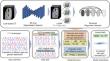
Methods: A deep-learning model developed based on 3312 low-dose chest computed tomography (LDCT) scans (trained with 2337 and tested with 975) achieved a mean dice similarity coefficient of 95.8% for T1-T12, L1, and L2 vertebral body (VB) segmentation on test data. We performed a model evaluation based on 4401 LDCT scans (obtained from scanners of 3 different manufacturers as external validation data). The BMD values of all individuals were extracted from three consecutive VBs: T12 to L2. Line regression and Bland‒Altman analyses were used to evaluate the overall detection performance. Sensitivity and specificity were used to evaluate the diagnostic performance for normal, osteopenia, and osteoporosis patients.
Results: Compared with the QCT results as the diagnostic standard, the BMD assessed had a mean error of (- 0.28, 2.37) mg/cm3. Overall, the sensitivity of a normal diagnosis was greater than that of a diagnosis of osteopenia or osteoporosis. For the diagnosis of osteoporosis, the model achieved a sensitivity > 86% and a specificity > 98%.
Conclusion: The developed tool is clinically applicable and helpful for the positioning and analysis of VBs, the measurement of BMD, and the screening of osteopenia and osteoporosis.
Clinical relevance statement: The developed system achieved high accuracy for automatic opportunistic osteoporosis screening using low-dose chest CT scans and performed well on CT images collected from different scanners.
Key points: Osteoporosis is a prevalent but underdiagnosed condition that can increase the risk of fractures. This system could automatically and opportunistically screen for osteoporosis using low-dose chest CT scans obtained for lung cancer screening. The developed system performed well on CT images collected from different scanners and did not differ with patient age or sex.
期刊介绍:
European Radiology (ER) continuously updates scientific knowledge in radiology by publication of strong original articles and state-of-the-art reviews written by leading radiologists. A well balanced combination of review articles, original papers, short communications from European radiological congresses and information on society matters makes ER an indispensable source for current information in this field.
This is the Journal of the European Society of Radiology, and the official journal of a number of societies.
From 2004-2008 supplements to European Radiology were published under its companion, European Radiology Supplements, ISSN 1613-3749.

 求助内容:
求助内容: 应助结果提醒方式:


