{"title":"伴有主动脉周和主动脉后左肾静脉的肾动脉瘘。","authors":"Jahira Banu, Nithya Dakshnamoorthy, Sulochana Sakthivel","doi":"10.1007/s12565-024-00796-0","DOIUrl":null,"url":null,"abstract":"<div><p>Variations of the left renal vein can be in the form of circumaortic vein or renal collar, retro-aortic vein, additional renal vein, or multiple primary tributaries. We report a unique complex venous pattern of concomitant circumaortic and retro-aortic left renal veins associated with a fenestrated left renal artery. Two renal veins, anterior and posterior to the renal artery, originated from the renal hilum. The anterior vein was further divided into two branches. One branch passed through the fenestrated renal artery to continue as the anterior limb of the circumaortic vein. It received the suprarenal and gonadal veins and drained into the inferior vena cava. The other branch coursed posterior to the fenestrated renal artery and joined the posterior renal vein. The posterior renal vein was divided into two branches: one forming the posterior limb of the circumaortic vein, and the other continued obliquely downwards as the retro-aortic vein. Variations in the left renal vein have been implicated in several clinical conditions, such as varicocele and pelvic varices. It also plays a crucial role in renal transplantation, as the left kidney is often used as the donor organ. Even though many reports have been published on circumaortic and retro-aortic veins, the complex venous pattern associated with a fenestrated renal artery has not been reported previously.</p></div>","PeriodicalId":7816,"journal":{"name":"Anatomical Science International","volume":"100 2","pages":"243 - 246"},"PeriodicalIF":1.7000,"publicationDate":"2024-09-03","publicationTypes":"Journal Article","fieldsOfStudy":null,"isOpenAccess":false,"openAccessPdf":"","citationCount":"0","resultStr":"{\"title\":\"Concomitant circumaortic and retro-aortic left renal veins associated with fenestrated renal artery\",\"authors\":\"Jahira Banu, Nithya Dakshnamoorthy, Sulochana Sakthivel\",\"doi\":\"10.1007/s12565-024-00796-0\",\"DOIUrl\":null,\"url\":null,\"abstract\":\"<div><p>Variations of the left renal vein can be in the form of circumaortic vein or renal collar, retro-aortic vein, additional renal vein, or multiple primary tributaries. We report a unique complex venous pattern of concomitant circumaortic and retro-aortic left renal veins associated with a fenestrated left renal artery. Two renal veins, anterior and posterior to the renal artery, originated from the renal hilum. The anterior vein was further divided into two branches. One branch passed through the fenestrated renal artery to continue as the anterior limb of the circumaortic vein. It received the suprarenal and gonadal veins and drained into the inferior vena cava. The other branch coursed posterior to the fenestrated renal artery and joined the posterior renal vein. The posterior renal vein was divided into two branches: one forming the posterior limb of the circumaortic vein, and the other continued obliquely downwards as the retro-aortic vein. Variations in the left renal vein have been implicated in several clinical conditions, such as varicocele and pelvic varices. It also plays a crucial role in renal transplantation, as the left kidney is often used as the donor organ. Even though many reports have been published on circumaortic and retro-aortic veins, the complex venous pattern associated with a fenestrated renal artery has not been reported previously.</p></div>\",\"PeriodicalId\":7816,\"journal\":{\"name\":\"Anatomical Science International\",\"volume\":\"100 2\",\"pages\":\"243 - 246\"},\"PeriodicalIF\":1.7000,\"publicationDate\":\"2024-09-03\",\"publicationTypes\":\"Journal Article\",\"fieldsOfStudy\":null,\"isOpenAccess\":false,\"openAccessPdf\":\"\",\"citationCount\":\"0\",\"resultStr\":null,\"platform\":\"Semanticscholar\",\"paperid\":null,\"PeriodicalName\":\"Anatomical Science International\",\"FirstCategoryId\":\"3\",\"ListUrlMain\":\"https://link.springer.com/article/10.1007/s12565-024-00796-0\",\"RegionNum\":4,\"RegionCategory\":\"医学\",\"ArticlePicture\":[],\"TitleCN\":null,\"AbstractTextCN\":null,\"PMCID\":null,\"EPubDate\":\"\",\"PubModel\":\"\",\"JCR\":\"Q3\",\"JCRName\":\"ANATOMY & MORPHOLOGY\",\"Score\":null,\"Total\":0}","platform":"Semanticscholar","paperid":null,"PeriodicalName":"Anatomical Science International","FirstCategoryId":"3","ListUrlMain":"https://link.springer.com/article/10.1007/s12565-024-00796-0","RegionNum":4,"RegionCategory":"医学","ArticlePicture":[],"TitleCN":null,"AbstractTextCN":null,"PMCID":null,"EPubDate":"","PubModel":"","JCR":"Q3","JCRName":"ANATOMY & MORPHOLOGY","Score":null,"Total":0}
Concomitant circumaortic and retro-aortic left renal veins associated with fenestrated renal artery
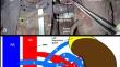
Variations of the left renal vein can be in the form of circumaortic vein or renal collar, retro-aortic vein, additional renal vein, or multiple primary tributaries. We report a unique complex venous pattern of concomitant circumaortic and retro-aortic left renal veins associated with a fenestrated left renal artery. Two renal veins, anterior and posterior to the renal artery, originated from the renal hilum. The anterior vein was further divided into two branches. One branch passed through the fenestrated renal artery to continue as the anterior limb of the circumaortic vein. It received the suprarenal and gonadal veins and drained into the inferior vena cava. The other branch coursed posterior to the fenestrated renal artery and joined the posterior renal vein. The posterior renal vein was divided into two branches: one forming the posterior limb of the circumaortic vein, and the other continued obliquely downwards as the retro-aortic vein. Variations in the left renal vein have been implicated in several clinical conditions, such as varicocele and pelvic varices. It also plays a crucial role in renal transplantation, as the left kidney is often used as the donor organ. Even though many reports have been published on circumaortic and retro-aortic veins, the complex venous pattern associated with a fenestrated renal artery has not been reported previously.
期刊介绍:
The official English journal of the Japanese Association of Anatomists, Anatomical Science International (formerly titled Kaibogaku Zasshi) publishes original research articles dealing with morphological sciences.
Coverage in the journal includes molecular, cellular, histological and gross anatomical studies on humans and on normal and experimental animals, as well as functional morphological, biochemical, physiological and behavioral studies if they include morphological analysis.

 求助内容:
求助内容: 应助结果提醒方式:


