Kemal Bugra Memis , Hasan Bulut , Hasibe Gokce Cinar , Berna Ucan , Cigdem Uner , Ozkan Kaya , Sonay Aydin
{"title":"通过胸部 CT 血管造影评估先天性心脏病患者腹腔干和肝动脉的变化","authors":"Kemal Bugra Memis , Hasan Bulut , Hasibe Gokce Cinar , Berna Ucan , Cigdem Uner , Ozkan Kaya , Sonay Aydin","doi":"10.1016/j.anpedi.2024.05.017","DOIUrl":null,"url":null,"abstract":"<div><h3>Introduction</h3><p>Understanding the variations of abdominal vascular structures is important for preventing complications of abdominal surgical procedures for gastrointestinal disease such as necrotizing enterocolitis or others that may arise in patients with congenital cardiac disease. We analysed the coeliac trunk and its branches in children with congenital heart disease to determine whether there is a greater prevalence of associated vascular abnormalities.</p></div><div><h3>Methods</h3><p>We retrospectively analysed thoracic computed tomography (CT) angiograms performed in our hospital in paediatric patients with congenital heart disease. We documented the anatomical variations observed in abdominal sections in which the coeliac trunk and hepatic arteries were included in the field of view. We used the Uflacker classification to describe anatomical variants of the coeliac trunk, and the Michels classification and its modified version (Hiatt classification) to describe the anatomy of the hepatic artery system.</p></div><div><h3>Results</h3><p>Our study included 178 patients with congenital heart disease. We identified coeliac trunk variants in 10.7% of the patients. Gastrosplenic trunk was to the most prevalent variant, amounting to 5.6% of total cases. We found hepatic artery variations in 19.1% of the patients. According to the Michels classification, the prevalence of accessory left hepatic artery arising from the left gastric artery as 4.5%, compared to 6.7% based on the Hiatt classification.</p></div><div><h3>Conclusion</h3><p>The prevalence of coeliac trunk and hepatic artery variations in patients with congenital heart disease was not greater in our study compared to other series in the literature. Clinicians must be vigilant about the variations detected in multislice CT scans to avoid complications resulting from vascular abnormalities, especially in patients who undergo abdominal surgery.</p></div>","PeriodicalId":7783,"journal":{"name":"Anales de pediatria","volume":"101 3","pages":"Pages 165-171"},"PeriodicalIF":1.5000,"publicationDate":"2024-09-01","publicationTypes":"Journal Article","fieldsOfStudy":null,"isOpenAccess":false,"openAccessPdf":"https://www.sciencedirect.com/science/article/pii/S1695403324001589/pdfft?md5=969721e9691bd11e63bcf3ef93707f99&pid=1-s2.0-S1695403324001589-main.pdf","citationCount":"0","resultStr":"{\"title\":\"Evaluación de las variaciones del tronco celíaco y la arteria hepática en la angiotomografía computarizada de tórax en los pacientes con cardiopatía congénita\",\"authors\":\"Kemal Bugra Memis , Hasan Bulut , Hasibe Gokce Cinar , Berna Ucan , Cigdem Uner , Ozkan Kaya , Sonay Aydin\",\"doi\":\"10.1016/j.anpedi.2024.05.017\",\"DOIUrl\":null,\"url\":null,\"abstract\":\"<div><h3>Introduction</h3><p>Understanding the variations of abdominal vascular structures is important for preventing complications of abdominal surgical procedures for gastrointestinal disease such as necrotizing enterocolitis or others that may arise in patients with congenital cardiac disease. We analysed the coeliac trunk and its branches in children with congenital heart disease to determine whether there is a greater prevalence of associated vascular abnormalities.</p></div><div><h3>Methods</h3><p>We retrospectively analysed thoracic computed tomography (CT) angiograms performed in our hospital in paediatric patients with congenital heart disease. We documented the anatomical variations observed in abdominal sections in which the coeliac trunk and hepatic arteries were included in the field of view. We used the Uflacker classification to describe anatomical variants of the coeliac trunk, and the Michels classification and its modified version (Hiatt classification) to describe the anatomy of the hepatic artery system.</p></div><div><h3>Results</h3><p>Our study included 178 patients with congenital heart disease. We identified coeliac trunk variants in 10.7% of the patients. Gastrosplenic trunk was to the most prevalent variant, amounting to 5.6% of total cases. We found hepatic artery variations in 19.1% of the patients. According to the Michels classification, the prevalence of accessory left hepatic artery arising from the left gastric artery as 4.5%, compared to 6.7% based on the Hiatt classification.</p></div><div><h3>Conclusion</h3><p>The prevalence of coeliac trunk and hepatic artery variations in patients with congenital heart disease was not greater in our study compared to other series in the literature. Clinicians must be vigilant about the variations detected in multislice CT scans to avoid complications resulting from vascular abnormalities, especially in patients who undergo abdominal surgery.</p></div>\",\"PeriodicalId\":7783,\"journal\":{\"name\":\"Anales de pediatria\",\"volume\":\"101 3\",\"pages\":\"Pages 165-171\"},\"PeriodicalIF\":1.5000,\"publicationDate\":\"2024-09-01\",\"publicationTypes\":\"Journal Article\",\"fieldsOfStudy\":null,\"isOpenAccess\":false,\"openAccessPdf\":\"https://www.sciencedirect.com/science/article/pii/S1695403324001589/pdfft?md5=969721e9691bd11e63bcf3ef93707f99&pid=1-s2.0-S1695403324001589-main.pdf\",\"citationCount\":\"0\",\"resultStr\":null,\"platform\":\"Semanticscholar\",\"paperid\":null,\"PeriodicalName\":\"Anales de pediatria\",\"FirstCategoryId\":\"3\",\"ListUrlMain\":\"https://www.sciencedirect.com/science/article/pii/S1695403324001589\",\"RegionNum\":4,\"RegionCategory\":\"医学\",\"ArticlePicture\":[],\"TitleCN\":null,\"AbstractTextCN\":null,\"PMCID\":null,\"EPubDate\":\"\",\"PubModel\":\"\",\"JCR\":\"Q2\",\"JCRName\":\"PEDIATRICS\",\"Score\":null,\"Total\":0}","platform":"Semanticscholar","paperid":null,"PeriodicalName":"Anales de pediatria","FirstCategoryId":"3","ListUrlMain":"https://www.sciencedirect.com/science/article/pii/S1695403324001589","RegionNum":4,"RegionCategory":"医学","ArticlePicture":[],"TitleCN":null,"AbstractTextCN":null,"PMCID":null,"EPubDate":"","PubModel":"","JCR":"Q2","JCRName":"PEDIATRICS","Score":null,"Total":0}
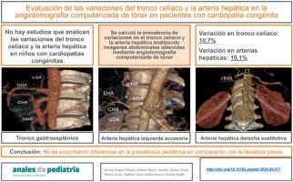
Evaluación de las variaciones del tronco celíaco y la arteria hepática en la angiotomografía computarizada de tórax en los pacientes con cardiopatía congénita
Introduction
Understanding the variations of abdominal vascular structures is important for preventing complications of abdominal surgical procedures for gastrointestinal disease such as necrotizing enterocolitis or others that may arise in patients with congenital cardiac disease. We analysed the coeliac trunk and its branches in children with congenital heart disease to determine whether there is a greater prevalence of associated vascular abnormalities.
Methods
We retrospectively analysed thoracic computed tomography (CT) angiograms performed in our hospital in paediatric patients with congenital heart disease. We documented the anatomical variations observed in abdominal sections in which the coeliac trunk and hepatic arteries were included in the field of view. We used the Uflacker classification to describe anatomical variants of the coeliac trunk, and the Michels classification and its modified version (Hiatt classification) to describe the anatomy of the hepatic artery system.
Results
Our study included 178 patients with congenital heart disease. We identified coeliac trunk variants in 10.7% of the patients. Gastrosplenic trunk was to the most prevalent variant, amounting to 5.6% of total cases. We found hepatic artery variations in 19.1% of the patients. According to the Michels classification, the prevalence of accessory left hepatic artery arising from the left gastric artery as 4.5%, compared to 6.7% based on the Hiatt classification.
Conclusion
The prevalence of coeliac trunk and hepatic artery variations in patients with congenital heart disease was not greater in our study compared to other series in the literature. Clinicians must be vigilant about the variations detected in multislice CT scans to avoid complications resulting from vascular abnormalities, especially in patients who undergo abdominal surgery.
期刊介绍:
La Asociación Española de Pediatría tiene como uno de sus objetivos principales la difusión de información científica rigurosa y actualizada sobre las distintas áreas de la pediatría. Anales de Pediatría es el Órgano de Expresión Científica de la Asociación y constituye el vehículo a través del cual se comunican los asociados. Publica trabajos originales sobre investigación clínica en pediatría procedentes de España y países latinoamericanos, así como artículos de revisión elaborados por los mejores profesionales de cada especialidad, las comunicaciones del congreso anual y los libros de actas de la Asociación, y guías de actuación elaboradas por las diferentes Sociedades/Secciones Especializadas integradas en la Asociación Española de Pediatría.

 求助内容:
求助内容: 应助结果提醒方式:


