{"title":"巨细胞病毒视网膜炎伴有高眼压葡萄膜炎掩盖的全视网膜闭塞性血管病变:病例报告。","authors":"Seongyong Jeong","doi":"10.12701/jyms.2024.00584","DOIUrl":null,"url":null,"abstract":"<p><p>Cytomegalovirus (CMV) retinitis is a rare disease, and overlapping manifestations involving the anterior segment are extremely uncommon. We report a patient who initially presented with persistent corneal edema and was later diagnosed with CMV retinitis. A 72-year-old man with uncontrolled intraocular pressure (IOP) in his right eye visited a tertiary hospital. At initial presentation, the IOP was 36 mmHg and the fundus was not clear due to corneal edema. Spectral domain optical coherence tomography revealed paracentral acute middle maculopathy (PAMM). Panretinal obstructive vasculopathy was observed on ultra-widefield fluorescein angiography. Three weeks later, trabeculectomy was performed to resolve the persistently high IOP. Once corneal edema improved, a white patch-like peripheral lesion and silver wire-like retinal vasculature were observed. Polymerase chain reaction of the aqueous humor was positive for CMV. Oral valganciclovir and intravitreal ganciclovir were administered as antiviral therapies. Despite treatment for 4 months, the final visual acuity was no light perception, with persistent corneal edema and neovascularization of the iris. We describe a rare case of the simultaneous occurrence of hypertensive uveitis and CMV retinitis. The presence of PAMM could be an initial identifiable sign of CMV retinitis, even in the presence of media opacity.</p>","PeriodicalId":74020,"journal":{"name":"Journal of Yeungnam medical science","volume":" ","pages":"300-305"},"PeriodicalIF":1.4000,"publicationDate":"2024-10-01","publicationTypes":"Journal Article","fieldsOfStudy":null,"isOpenAccess":false,"openAccessPdf":"https://www.ncbi.nlm.nih.gov/pmc/articles/PMC11534412/pdf/","citationCount":"0","resultStr":"{\"title\":\"Cytomegalovirus retinitis with panretinal occlusive vasculopathy concealed by hypertensive uveitis: a case report.\",\"authors\":\"Seongyong Jeong\",\"doi\":\"10.12701/jyms.2024.00584\",\"DOIUrl\":null,\"url\":null,\"abstract\":\"<p><p>Cytomegalovirus (CMV) retinitis is a rare disease, and overlapping manifestations involving the anterior segment are extremely uncommon. We report a patient who initially presented with persistent corneal edema and was later diagnosed with CMV retinitis. A 72-year-old man with uncontrolled intraocular pressure (IOP) in his right eye visited a tertiary hospital. At initial presentation, the IOP was 36 mmHg and the fundus was not clear due to corneal edema. Spectral domain optical coherence tomography revealed paracentral acute middle maculopathy (PAMM). Panretinal obstructive vasculopathy was observed on ultra-widefield fluorescein angiography. Three weeks later, trabeculectomy was performed to resolve the persistently high IOP. Once corneal edema improved, a white patch-like peripheral lesion and silver wire-like retinal vasculature were observed. Polymerase chain reaction of the aqueous humor was positive for CMV. Oral valganciclovir and intravitreal ganciclovir were administered as antiviral therapies. Despite treatment for 4 months, the final visual acuity was no light perception, with persistent corneal edema and neovascularization of the iris. We describe a rare case of the simultaneous occurrence of hypertensive uveitis and CMV retinitis. The presence of PAMM could be an initial identifiable sign of CMV retinitis, even in the presence of media opacity.</p>\",\"PeriodicalId\":74020,\"journal\":{\"name\":\"Journal of Yeungnam medical science\",\"volume\":\" \",\"pages\":\"300-305\"},\"PeriodicalIF\":1.4000,\"publicationDate\":\"2024-10-01\",\"publicationTypes\":\"Journal Article\",\"fieldsOfStudy\":null,\"isOpenAccess\":false,\"openAccessPdf\":\"https://www.ncbi.nlm.nih.gov/pmc/articles/PMC11534412/pdf/\",\"citationCount\":\"0\",\"resultStr\":null,\"platform\":\"Semanticscholar\",\"paperid\":null,\"PeriodicalName\":\"Journal of Yeungnam medical science\",\"FirstCategoryId\":\"1085\",\"ListUrlMain\":\"https://doi.org/10.12701/jyms.2024.00584\",\"RegionNum\":0,\"RegionCategory\":null,\"ArticlePicture\":[],\"TitleCN\":null,\"AbstractTextCN\":null,\"PMCID\":null,\"EPubDate\":\"2024/8/30 0:00:00\",\"PubModel\":\"Epub\",\"JCR\":\"Q3\",\"JCRName\":\"MEDICINE, GENERAL & INTERNAL\",\"Score\":null,\"Total\":0}","platform":"Semanticscholar","paperid":null,"PeriodicalName":"Journal of Yeungnam medical science","FirstCategoryId":"1085","ListUrlMain":"https://doi.org/10.12701/jyms.2024.00584","RegionNum":0,"RegionCategory":null,"ArticlePicture":[],"TitleCN":null,"AbstractTextCN":null,"PMCID":null,"EPubDate":"2024/8/30 0:00:00","PubModel":"Epub","JCR":"Q3","JCRName":"MEDICINE, GENERAL & INTERNAL","Score":null,"Total":0}
Cytomegalovirus retinitis with panretinal occlusive vasculopathy concealed by hypertensive uveitis: a case report.
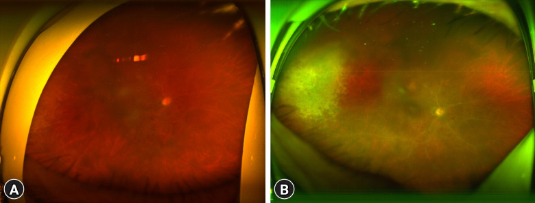
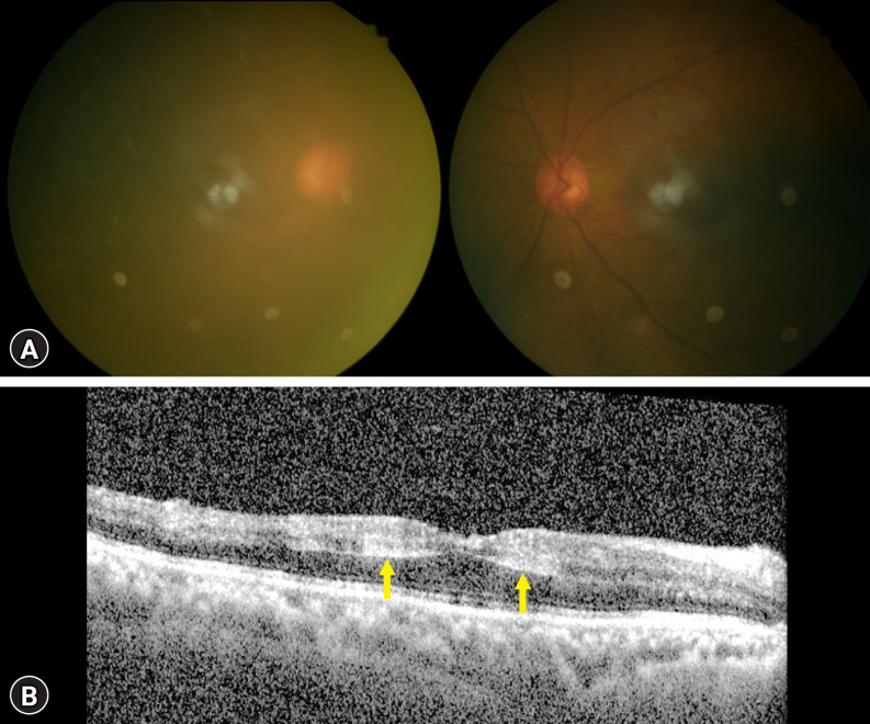
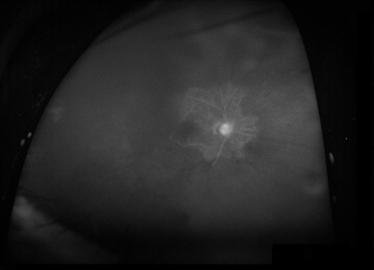
Cytomegalovirus (CMV) retinitis is a rare disease, and overlapping manifestations involving the anterior segment are extremely uncommon. We report a patient who initially presented with persistent corneal edema and was later diagnosed with CMV retinitis. A 72-year-old man with uncontrolled intraocular pressure (IOP) in his right eye visited a tertiary hospital. At initial presentation, the IOP was 36 mmHg and the fundus was not clear due to corneal edema. Spectral domain optical coherence tomography revealed paracentral acute middle maculopathy (PAMM). Panretinal obstructive vasculopathy was observed on ultra-widefield fluorescein angiography. Three weeks later, trabeculectomy was performed to resolve the persistently high IOP. Once corneal edema improved, a white patch-like peripheral lesion and silver wire-like retinal vasculature were observed. Polymerase chain reaction of the aqueous humor was positive for CMV. Oral valganciclovir and intravitreal ganciclovir were administered as antiviral therapies. Despite treatment for 4 months, the final visual acuity was no light perception, with persistent corneal edema and neovascularization of the iris. We describe a rare case of the simultaneous occurrence of hypertensive uveitis and CMV retinitis. The presence of PAMM could be an initial identifiable sign of CMV retinitis, even in the presence of media opacity.

 求助内容:
求助内容: 应助结果提醒方式:


