Quan Shi, Yang Huang, Na Huo, Yi Jiang, Tong Zhang, Juncheng Wang
{"title":"以修复为导向的下颌第一磨牙牙槽骨解剖分析及其对即刻种植手术的影响:一项 CBCT 研究。","authors":"Quan Shi, Yang Huang, Na Huo, Yi Jiang, Tong Zhang, Juncheng Wang","doi":"10.4047/jap.2024.16.4.212","DOIUrl":null,"url":null,"abstract":"<p><strong>Purpose: </strong>This cone-beam computed tomography (CBCT) study aimed to analyze the anatomical characteristics of alveolar bone at mandibular first molar (MFM) and their implications for immediate implant placement surgery.</p><p><strong>Materials and methods: </strong>100 patients with 140 MFMs were reviewed retrospectively. We first performed a 3D reconstruction of the patient's CBCT data to determine a reference plane with ideal implant placement and orientation. The following parameters of MFM region were analyzed: mesial-distal socket size (MD-SS), buccal-lingual socket size (BL-SS), root furcation fornix to inferior alveolar nerve (IAN) distance (RF-I), interradicular bone thickness (IRB), mesial/distal root apex to the IAN distance (MRA-I/DRA-I), thickness of the buccal/lingual bone of the mesial root (MR-B/MR-L), thickness of the buccal/lingual bone of the distal root (DR-B/DR-L).</p><p><strong>Results: </strong>The MD-SS of MFM was 8.74 ± 0.76 mm, and the BL-SS was 8.26 ± 0.72 mm. The MR-B, DR-B was 1.01 ± 0.40 mm and 1.14 ± 0.50 mm, and the difference was statistically significant (<i>P</i> = .001). The values of the MR-L, DR-L were 2.71 ± 0.78 mm and 3.09 ± 0.73 mm, and the difference was also statistically significant (<i>P</i> < .001). The mean distance of RF-I was 15.68 ± 2.13 mm, and the MRA-I was 7.06 ± 2.22 mm, which was greater than that of DRA-I (6.48 ± 2.30 mm, <i>P</i> < .001). The IRB at 2 mm, 4 mm apical from the furcation fornix, and at apex level was 2.81 ± 0.50 mm, 3.30 ± 0.62 mm, and 4.44 ± 1.02 mm, respectively.</p><p><strong>Conclusion: </strong>There is relatively sufficient bone mass in interradicular bone in height, but an adequate width is lacking for the bone between the mesial and distal root after the extraction of the MFM for immediate implantation. The thickness of the MFM buccal bone is relative thin, especially for the mesial root.</p>","PeriodicalId":51291,"journal":{"name":"Journal of Advanced Prosthodontics","volume":"16 4","pages":"212-220"},"PeriodicalIF":2.5000,"publicationDate":"2024-08-01","publicationTypes":"Journal Article","fieldsOfStudy":null,"isOpenAccess":false,"openAccessPdf":"https://www.ncbi.nlm.nih.gov/pmc/articles/PMC11361819/pdf/","citationCount":"0","resultStr":"{\"title\":\"Restoration-oriented anatomical analysis of alveolar bone at mandibular first molars and implications for immediate implant placement surgery: a CBCT study.\",\"authors\":\"Quan Shi, Yang Huang, Na Huo, Yi Jiang, Tong Zhang, Juncheng Wang\",\"doi\":\"10.4047/jap.2024.16.4.212\",\"DOIUrl\":null,\"url\":null,\"abstract\":\"<p><strong>Purpose: </strong>This cone-beam computed tomography (CBCT) study aimed to analyze the anatomical characteristics of alveolar bone at mandibular first molar (MFM) and their implications for immediate implant placement surgery.</p><p><strong>Materials and methods: </strong>100 patients with 140 MFMs were reviewed retrospectively. We first performed a 3D reconstruction of the patient's CBCT data to determine a reference plane with ideal implant placement and orientation. The following parameters of MFM region were analyzed: mesial-distal socket size (MD-SS), buccal-lingual socket size (BL-SS), root furcation fornix to inferior alveolar nerve (IAN) distance (RF-I), interradicular bone thickness (IRB), mesial/distal root apex to the IAN distance (MRA-I/DRA-I), thickness of the buccal/lingual bone of the mesial root (MR-B/MR-L), thickness of the buccal/lingual bone of the distal root (DR-B/DR-L).</p><p><strong>Results: </strong>The MD-SS of MFM was 8.74 ± 0.76 mm, and the BL-SS was 8.26 ± 0.72 mm. The MR-B, DR-B was 1.01 ± 0.40 mm and 1.14 ± 0.50 mm, and the difference was statistically significant (<i>P</i> = .001). The values of the MR-L, DR-L were 2.71 ± 0.78 mm and 3.09 ± 0.73 mm, and the difference was also statistically significant (<i>P</i> < .001). The mean distance of RF-I was 15.68 ± 2.13 mm, and the MRA-I was 7.06 ± 2.22 mm, which was greater than that of DRA-I (6.48 ± 2.30 mm, <i>P</i> < .001). The IRB at 2 mm, 4 mm apical from the furcation fornix, and at apex level was 2.81 ± 0.50 mm, 3.30 ± 0.62 mm, and 4.44 ± 1.02 mm, respectively.</p><p><strong>Conclusion: </strong>There is relatively sufficient bone mass in interradicular bone in height, but an adequate width is lacking for the bone between the mesial and distal root after the extraction of the MFM for immediate implantation. The thickness of the MFM buccal bone is relative thin, especially for the mesial root.</p>\",\"PeriodicalId\":51291,\"journal\":{\"name\":\"Journal of Advanced Prosthodontics\",\"volume\":\"16 4\",\"pages\":\"212-220\"},\"PeriodicalIF\":2.5000,\"publicationDate\":\"2024-08-01\",\"publicationTypes\":\"Journal Article\",\"fieldsOfStudy\":null,\"isOpenAccess\":false,\"openAccessPdf\":\"https://www.ncbi.nlm.nih.gov/pmc/articles/PMC11361819/pdf/\",\"citationCount\":\"0\",\"resultStr\":null,\"platform\":\"Semanticscholar\",\"paperid\":null,\"PeriodicalName\":\"Journal of Advanced Prosthodontics\",\"FirstCategoryId\":\"3\",\"ListUrlMain\":\"https://doi.org/10.4047/jap.2024.16.4.212\",\"RegionNum\":3,\"RegionCategory\":\"医学\",\"ArticlePicture\":[],\"TitleCN\":null,\"AbstractTextCN\":null,\"PMCID\":null,\"EPubDate\":\"2024/8/20 0:00:00\",\"PubModel\":\"Epub\",\"JCR\":\"Q1\",\"JCRName\":\"DENTISTRY, ORAL SURGERY & MEDICINE\",\"Score\":null,\"Total\":0}","platform":"Semanticscholar","paperid":null,"PeriodicalName":"Journal of Advanced Prosthodontics","FirstCategoryId":"3","ListUrlMain":"https://doi.org/10.4047/jap.2024.16.4.212","RegionNum":3,"RegionCategory":"医学","ArticlePicture":[],"TitleCN":null,"AbstractTextCN":null,"PMCID":null,"EPubDate":"2024/8/20 0:00:00","PubModel":"Epub","JCR":"Q1","JCRName":"DENTISTRY, ORAL SURGERY & MEDICINE","Score":null,"Total":0}
Restoration-oriented anatomical analysis of alveolar bone at mandibular first molars and implications for immediate implant placement surgery: a CBCT study.
Purpose: This cone-beam computed tomography (CBCT) study aimed to analyze the anatomical characteristics of alveolar bone at mandibular first molar (MFM) and their implications for immediate implant placement surgery.
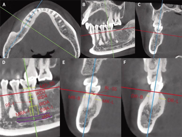
Materials and methods: 100 patients with 140 MFMs were reviewed retrospectively. We first performed a 3D reconstruction of the patient's CBCT data to determine a reference plane with ideal implant placement and orientation. The following parameters of MFM region were analyzed: mesial-distal socket size (MD-SS), buccal-lingual socket size (BL-SS), root furcation fornix to inferior alveolar nerve (IAN) distance (RF-I), interradicular bone thickness (IRB), mesial/distal root apex to the IAN distance (MRA-I/DRA-I), thickness of the buccal/lingual bone of the mesial root (MR-B/MR-L), thickness of the buccal/lingual bone of the distal root (DR-B/DR-L).
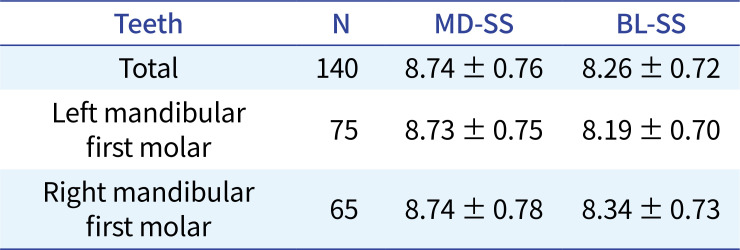
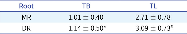
Results: The MD-SS of MFM was 8.74 ± 0.76 mm, and the BL-SS was 8.26 ± 0.72 mm. The MR-B, DR-B was 1.01 ± 0.40 mm and 1.14 ± 0.50 mm, and the difference was statistically significant (P = .001). The values of the MR-L, DR-L were 2.71 ± 0.78 mm and 3.09 ± 0.73 mm, and the difference was also statistically significant (P < .001). The mean distance of RF-I was 15.68 ± 2.13 mm, and the MRA-I was 7.06 ± 2.22 mm, which was greater than that of DRA-I (6.48 ± 2.30 mm, P < .001). The IRB at 2 mm, 4 mm apical from the furcation fornix, and at apex level was 2.81 ± 0.50 mm, 3.30 ± 0.62 mm, and 4.44 ± 1.02 mm, respectively.
Conclusion: There is relatively sufficient bone mass in interradicular bone in height, but an adequate width is lacking for the bone between the mesial and distal root after the extraction of the MFM for immediate implantation. The thickness of the MFM buccal bone is relative thin, especially for the mesial root.
期刊介绍:
This journal aims to convey scientific and clinical progress in the field of prosthodontics and its related areas to many dental communities concerned with esthetic and functional restorations, occlusion, implants, prostheses, and biomaterials related to prosthodontics.
This journal publishes
• Original research data of high scientific merit in the field of diagnosis, function, esthetics and stomatognathic physiology related to prosthodontic rehabilitation, physiology and mechanics of occlusion, mechanical and biologic aspects of prosthodontic materials including dental implants.
• Review articles by experts on controversies and new developments in prosthodontics.
• Case reports if they provide or document new fundamental knowledge.
 求助内容:
求助内容: 应助结果提醒方式:


