Giacomo Valli, Rui Wu, Dean Minnock, Giuseppe Sirago, Giosuè Annibalini, Andrea Casolo, Alessandro Del Vecchio, Luana Toniolo, Elena Barbieri, Giuseppe De Vito
{"title":"非侵入性运动单元分析能否揭示无并发症的 1 型糖尿病患者的不同发力神经策略?","authors":"Giacomo Valli, Rui Wu, Dean Minnock, Giuseppe Sirago, Giosuè Annibalini, Andrea Casolo, Alessandro Del Vecchio, Luana Toniolo, Elena Barbieri, Giuseppe De Vito","doi":"10.1007/s00421-024-05595-z","DOIUrl":null,"url":null,"abstract":"<p><strong>Purpose: </strong>to investigate the early consequences of type 1 diabetes (T1D) on the neural strategies of muscle force production.</p><p><strong>Methods: </strong>motor unit (MU) activity was recorded from the vastus lateralis muscle with High-Density surface Electromyography during isometric knee extension at 20 and 40% of maximum voluntary contraction (MVC) in 8 T1D (4 males, 4 females, 30.5 ± 3.6 years) and 8 matched control (4 males, 4 females, 27.3 ± 5.9 years) participants. Muscle biopsies were also collected from vastus lateralis for fiber type analysis, including myosin heavy chain (MyHC) isoform content via protein and mRNA expression.</p><p><strong>Results: </strong>MVC was comparable between groups as well as MU conduction velocity, action potentials' amplitude and proportions of MyHC protein isoforms. Nonetheless, MU discharge rate, relative derecruitment thresholds and mRNA expression of MyHC isoform I were lower in T1D.</p><p><strong>Conclusions: </strong>young people with uncomplicated T1D present a different neural control of muscle force production. Furthermore, differences are detectable non-invasively in absence of any functional manifestation (i.e., force production and fiber type distribution). These novel findings suggest that T1D has early consequences on the neuromuscular system and highlights the necessity of a better characterization of neural control in this population.</p>","PeriodicalId":12005,"journal":{"name":"European Journal of Applied Physiology","volume":" ","pages":"247-259"},"PeriodicalIF":2.8000,"publicationDate":"2025-01-01","publicationTypes":"Journal Article","fieldsOfStudy":null,"isOpenAccess":false,"openAccessPdf":"","citationCount":"0","resultStr":"{\"title\":\"Can non-invasive motor unit analysis reveal distinct neural strategies of force production in young with uncomplicated type 1 diabetes?\",\"authors\":\"Giacomo Valli, Rui Wu, Dean Minnock, Giuseppe Sirago, Giosuè Annibalini, Andrea Casolo, Alessandro Del Vecchio, Luana Toniolo, Elena Barbieri, Giuseppe De Vito\",\"doi\":\"10.1007/s00421-024-05595-z\",\"DOIUrl\":null,\"url\":null,\"abstract\":\"<p><strong>Purpose: </strong>to investigate the early consequences of type 1 diabetes (T1D) on the neural strategies of muscle force production.</p><p><strong>Methods: </strong>motor unit (MU) activity was recorded from the vastus lateralis muscle with High-Density surface Electromyography during isometric knee extension at 20 and 40% of maximum voluntary contraction (MVC) in 8 T1D (4 males, 4 females, 30.5 ± 3.6 years) and 8 matched control (4 males, 4 females, 27.3 ± 5.9 years) participants. Muscle biopsies were also collected from vastus lateralis for fiber type analysis, including myosin heavy chain (MyHC) isoform content via protein and mRNA expression.</p><p><strong>Results: </strong>MVC was comparable between groups as well as MU conduction velocity, action potentials' amplitude and proportions of MyHC protein isoforms. Nonetheless, MU discharge rate, relative derecruitment thresholds and mRNA expression of MyHC isoform I were lower in T1D.</p><p><strong>Conclusions: </strong>young people with uncomplicated T1D present a different neural control of muscle force production. Furthermore, differences are detectable non-invasively in absence of any functional manifestation (i.e., force production and fiber type distribution). These novel findings suggest that T1D has early consequences on the neuromuscular system and highlights the necessity of a better characterization of neural control in this population.</p>\",\"PeriodicalId\":12005,\"journal\":{\"name\":\"European Journal of Applied Physiology\",\"volume\":\" \",\"pages\":\"247-259\"},\"PeriodicalIF\":2.8000,\"publicationDate\":\"2025-01-01\",\"publicationTypes\":\"Journal Article\",\"fieldsOfStudy\":null,\"isOpenAccess\":false,\"openAccessPdf\":\"\",\"citationCount\":\"0\",\"resultStr\":null,\"platform\":\"Semanticscholar\",\"paperid\":null,\"PeriodicalName\":\"European Journal of Applied Physiology\",\"FirstCategoryId\":\"3\",\"ListUrlMain\":\"https://doi.org/10.1007/s00421-024-05595-z\",\"RegionNum\":3,\"RegionCategory\":\"医学\",\"ArticlePicture\":[],\"TitleCN\":null,\"AbstractTextCN\":null,\"PMCID\":null,\"EPubDate\":\"2024/8/30 0:00:00\",\"PubModel\":\"Epub\",\"JCR\":\"Q2\",\"JCRName\":\"PHYSIOLOGY\",\"Score\":null,\"Total\":0}","platform":"Semanticscholar","paperid":null,"PeriodicalName":"European Journal of Applied Physiology","FirstCategoryId":"3","ListUrlMain":"https://doi.org/10.1007/s00421-024-05595-z","RegionNum":3,"RegionCategory":"医学","ArticlePicture":[],"TitleCN":null,"AbstractTextCN":null,"PMCID":null,"EPubDate":"2024/8/30 0:00:00","PubModel":"Epub","JCR":"Q2","JCRName":"PHYSIOLOGY","Score":null,"Total":0}
Can non-invasive motor unit analysis reveal distinct neural strategies of force production in young with uncomplicated type 1 diabetes?
Purpose: to investigate the early consequences of type 1 diabetes (T1D) on the neural strategies of muscle force production.
Methods: motor unit (MU) activity was recorded from the vastus lateralis muscle with High-Density surface Electromyography during isometric knee extension at 20 and 40% of maximum voluntary contraction (MVC) in 8 T1D (4 males, 4 females, 30.5 ± 3.6 years) and 8 matched control (4 males, 4 females, 27.3 ± 5.9 years) participants. Muscle biopsies were also collected from vastus lateralis for fiber type analysis, including myosin heavy chain (MyHC) isoform content via protein and mRNA expression.
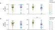
Results: MVC was comparable between groups as well as MU conduction velocity, action potentials' amplitude and proportions of MyHC protein isoforms. Nonetheless, MU discharge rate, relative derecruitment thresholds and mRNA expression of MyHC isoform I were lower in T1D.
Conclusions: young people with uncomplicated T1D present a different neural control of muscle force production. Furthermore, differences are detectable non-invasively in absence of any functional manifestation (i.e., force production and fiber type distribution). These novel findings suggest that T1D has early consequences on the neuromuscular system and highlights the necessity of a better characterization of neural control in this population.
期刊介绍:
The European Journal of Applied Physiology (EJAP) aims to promote mechanistic advances in human integrative and translational physiology. Physiology is viewed broadly, having overlapping context with related disciplines such as biomechanics, biochemistry, endocrinology, ergonomics, immunology, motor control, and nutrition. EJAP welcomes studies dealing with physical exercise, training and performance. Studies addressing physiological mechanisms are preferred over descriptive studies. Papers dealing with animal models or pathophysiological conditions are not excluded from consideration, but must be clearly relevant to human physiology.

 求助内容:
求助内容: 应助结果提醒方式:


